
|
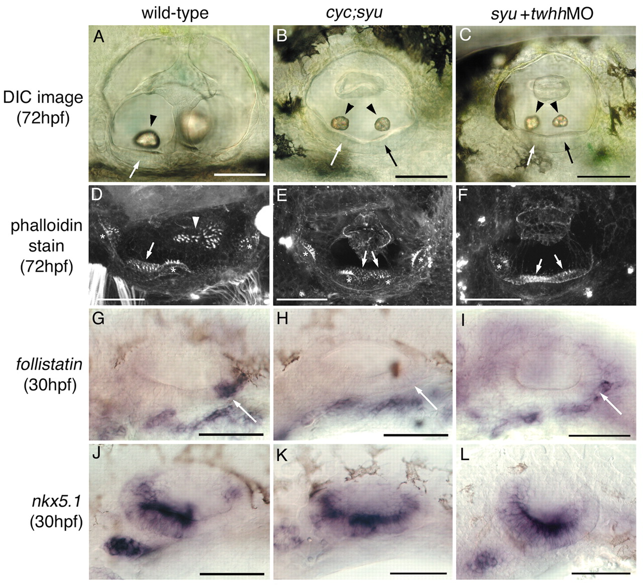
Fig. 7 Anteriorised ear phenotypes in cyc;syu double mutants and twhh antisense morpholino-injected syut4 embryos. Lateral views; anterior towards the left, dorsal towards the top. (A,D,G,J) Wild-type ear pattern; A is taken from Fig. 3 for comparison. (B,E,H,K) Ears of syu;cyc double mutants. (C,F,I,L) Ears of syut4 mutant embryos injected with 0.25mM twhh morpholino. (A-C) DIC images of live ears. The ears of cyc;syu mutants and twhh MO-injected embryos have two small, lateral otoliths (arrowheads B,C) resembling the anterior otolith of ears from wild-type embryos (arrowhead, A). Thickened sensory epithelium is present at both the anterior and the posterior of the vesicle in cyc;syu and syu +twhh MO embryos (arrows, B,C) rather than just at the anterior as in the wild-type (arrow, A). (D-F) Confocal images of FITC-phalloidin stains. Hair cells on the ventral floor are present at both the posterior and anterior of the vesicle in cyc;syu and syu +twhh MO embryos (arrows, E,F) rather than just at the anterior as in the wild-type (arrow, D). The posterior macula (arrowhead, D) is missing from the medial wall in cyc;syu and syu + twhh MO ears (E,F). Four cristae (*) rather than the usual three are present in some cyc;syu and syu + twhh MO embryos (e.g. E). Three cristae were present in the ear shown in F, but only one is in the focal plane. (G-L) In situ hybridisation. Arrows indicate the posterior domain of follistatin expression in the wild type (G). This is absent in cyc;syu and syu+twhh MO ears (H,I). Anterior nkx5.1 expression (J) is expanded in cyc;syu and (less extensively) in syu+twhh MO ears (K,L). Scale bars: 50 μm.