
Image
|
Figure Caption
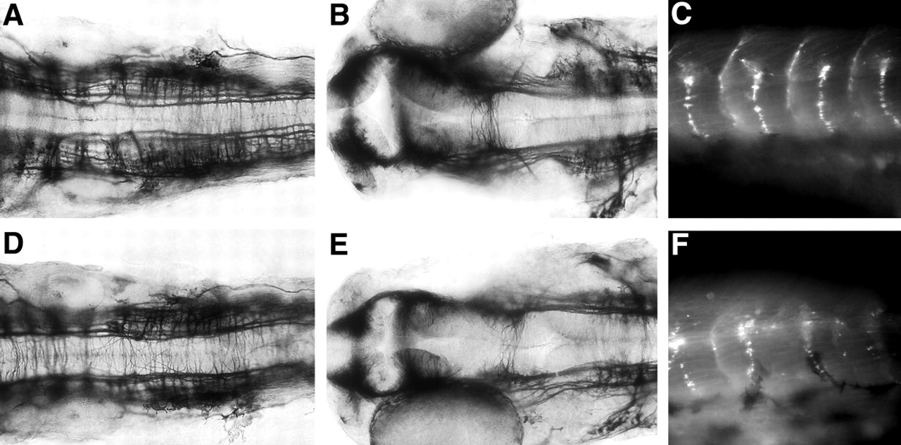
Fig. 5 CNS and neuromuscular junction organisation is unaffected in dystroglycan morphants. Acetylated tubulin staining shows the pattern of axonal scaffolds in control MO-injected (A,B) and dystroglycan morphant (D,E) embryos. (A,D) Hindbrain views. (B,E) Anterior views. We could find no consistent morphological difference between control MO-injected (C) and dystroglycan morphant (F) after staining for AChRs. All embryos are at 48 hpf.
Figure Data
Acknowledgments
This image is the copyrighted work of the attributed author or publisher, and
ZFIN has permission only to display this image to its users.
Additional permissions should be obtained from the applicable author or publisher of the image.
Full text @ Development