
Image
|
Figure Caption
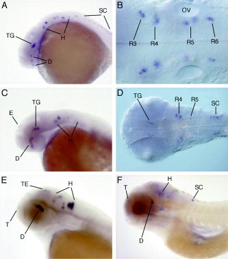
Fig. 5 Expression of the drd2b gene. A: A 24 hours postfertilization (hpf) embryo (lateral view). B: Differential interference contrast microscopy (DIC) image of a 24 hpf embryo. C: At 36 hpf (lateral view). D: DIC image of a 36 hpf embryo. E: At 48 hpf (lateral view). F: 5 dpf (lateral view). D, diencephalon; E, epiphysis; H, hindbrain; R, rhombomere; SC, spinal cord; T, telencephalon; TE, tectum; TG, tegmentum. The unstained otic vesicle (OV) is shown for orientation.
Figure Data
Acknowledgments
This image is the copyrighted work of the attributed author or publisher, and
ZFIN has permission only to display this image to its users.
Additional permissions should be obtained from the applicable author or publisher of the image.
Full text @ Dev. Dyn.