
Fig. S5
- ID
- ZDB-IMAGE-101007-35
- Publication
- Imai et al., 2010 - The ubiquitin proteasome system is required for cell proliferation of the lens epithelium and for differentiation of lens fiber cells in zebrafish
- All Figures
- Figures for Imai et al., 2010
|
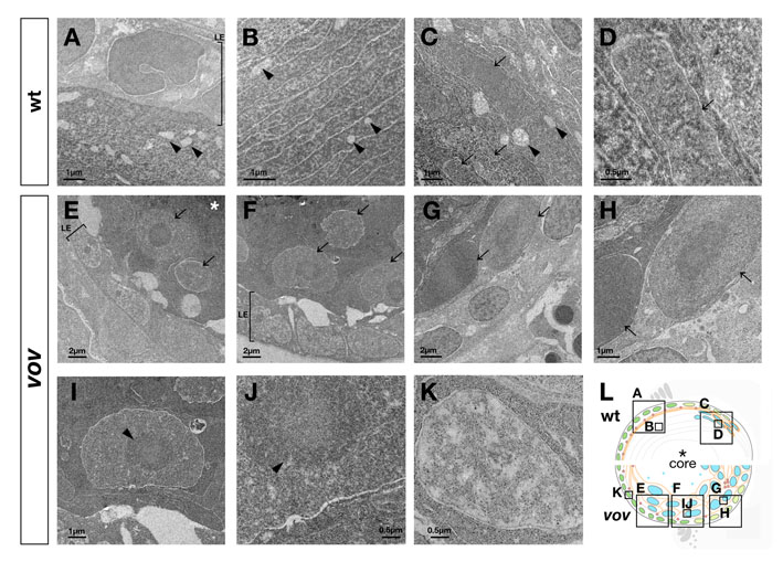
Fig. S5 Ultrastructure of the vov mutant lens. (A-D) Transmission electron microscopy (TEM) images of wild-type lens at 72 hpf. No nuclei of lens fiber cells were observed underneath the anterior lens epithelium (A). Arrowheads indicate subcellular structures similar to mitochondria in the lens fiber region underneath the anterior lens epithelium (arrowheads, A-C). In the posterior region of the lens sphere, flat nuclei of lens fiber cells are stacked regularly (arrows, C,D). Nuclei of lens fiber cells were electron-densely stained in patches (D). (E-K) TEM images of the vov mutant lens at 72 hpf. Large nuclei are observed underneath the anterior epithelium (arrows, E), near the equator (arrows, F) and in the posterior region of the lens sphere (arrows, G,H). Unlike in the wild type, most of the lens fiber nuclei were stained uniformly, and also contained a dark-stained circular region inside (arrowhead, I,J). In contrast to lens fiber cells, nuclei of lens epithelium were electron-densely stained in patches (K). (L) Schematic of wild-type and vov mutant lens. Boxes indicate areas shown in A-K. Asterisks indicate the lens fiber core, which is darkly stained in TEM analyses (A,C). LE, lens epithelium.