
Fig. 3
- ID
- ZDB-IMAGE-101007-28
- Genes
- Publication
- Imai et al., 2010 - The ubiquitin proteasome system is required for cell proliferation of the lens epithelium and for differentiation of lens fiber cells in zebrafish
- All Figures
- Figures for Imai et al., 2010
|
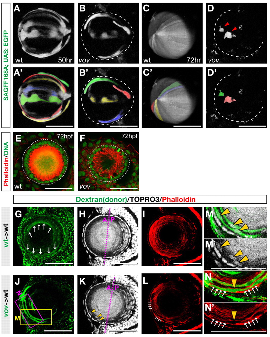
Fig. 3 Morphogenesis of lens fiber cells is affected in the vov mutant. (A-D2) Confocal images of the lens of 50 hpf wild type (A) and vov mutant (B) and 72 hpf wild type (C) and vov mutant (D) zebrafish expressing the SAGFF168A; UAS:EGFP transgenes. Individual lens fiber cells are indicated by pseudocolors (A2-D2). In the vov mutant, GFP-expressing cells do not maintain a fiber-like structure and appear to undergo degradation (D, arrowheads). (E,F) Anterior view of 72 hpf wild-type (E) and the vov mutant (F) lens labeled with Rhodamine-conjugated phalloidin (red) and Sytox Green (green). (G-N′) Transplantation of wild-type (G-I) and vov mutant (J-N) donor cells into wild-type recipient lens. Labeling is with biotin-dextran (green) (G,J), TOPRO3 (gray) (H,K) and phalloidin (red) (I,L). (M,N) High magnification of the boxed regions shown in J. Wild-type donor cells transplanted into wild-type recipient lens elongate to be very thin, resulting in weak signals (arrows, G). Nuclei of vov mutant donor lens fiber cells were frequently positioned in the anterior lens fiber region (yellow arrowheads, K,M,M2) and F-actin density is lower in the vov mutant (white arrows, L,N,N2). Scale bars: 50 µm.