
|
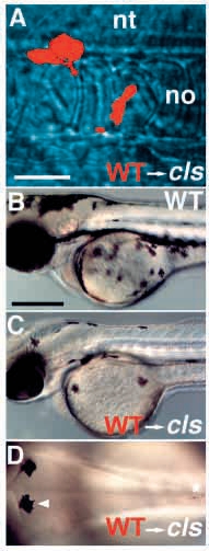
Fig. 9 Wild-type→cls- chimaeric embryos show enhanced numbers of pigment cells. Wild-type donor cells labeled with rhodamine dextran (red) and transplanted into an unlabeled cls- host embryo at shield stage frequently contributed to neural crest. Three cells are seen here migrating past the notochord (no) at 24 hpf (A). Some chimaeric cls- embryos show many (C) or few (D) melanophores by 2 dpf; all such embryos are clearly distinguishable from wild types (B). (D) All these melanophores have a wild-type appearance [compare wild-type melanophore (arrowhead) and mutant melanophore (asterisk)]. A-C, lateral views; D, dorsal view of posterior head. nt, neural tube. Scale bars, 50 μm (A) and 250 μm (B-D).