
Image
|
Figure Caption
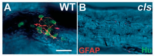
Fig. 7 GFAP-positive enteric glia are largely absent in cls- larvae. Dissected 5 dpf hindgut preparations stained with anti-Hu (green) and anti-GFAP (red) antibodies clearly reveal the enteric nervous system in the wild type (A) and its essential absence from cls- siblings (B). Confocal sections of the fluorescent labeling are superimposed on a DIC image of the gut. Scale bar, 20 μm.
Figure Data
Acknowledgments
This image is the copyrighted work of the attributed author or publisher, and
ZFIN has permission only to display this image to its users.
Additional permissions should be obtained from the applicable author or publisher of the image.
Full text @ Development