
|
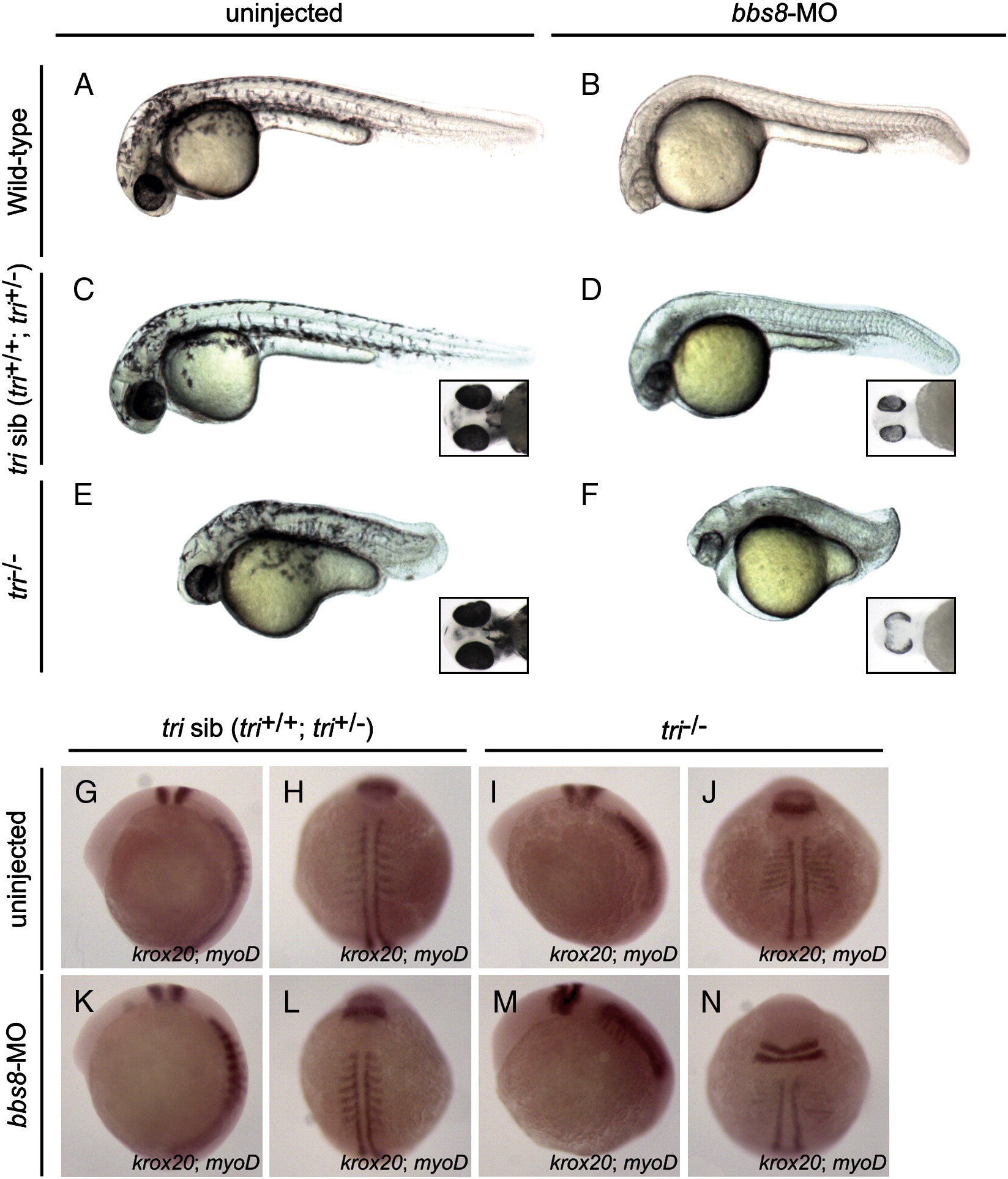
Fig. 1 Bbs8 interacts with Vangl2 during zebrafish development. Injection of bbs8-MO at the 1-2-cell stages into trilobite (tri) zebrafish. A-F) At 30 hpf, defective CE cell movement of tri-/- was enhanced after injection of bbs8-MO, resulting in severe body axis compression. Inset C-F) Ventral view of zebrafish embryos at 48 hpf. Note varied spacing between eyes. G-N) Visualisation of somites (myoD) and rhombomeres 3 and 5 (krox20) by in situ hybridisation at the 8-somite stage. Patterning is unaltered in tri siblings. bbs8-MO injected tri-/- embryos had more severe CE defects as evidenced by compressed somites and a wider presumptive neural tube, and a shorter distance between rhombomere 5 and the first somite. Views: lateral (A–G, K, I, M), dorsal (H, L, J, N).
Reprinted from Developmental Biology, 345(2), May-Simera, H.L., Kai, M., Hernandez, V., Osborn, D.P., Tada, M., and Beales, P.L., Bbs8, together with the planar cell polarity protein Vangl2, is required to establish left-right asymmetry in zebrafish, 215-225, Copyright (2010) with permission from Elsevier. Full text @ Dev. Biol.