
|
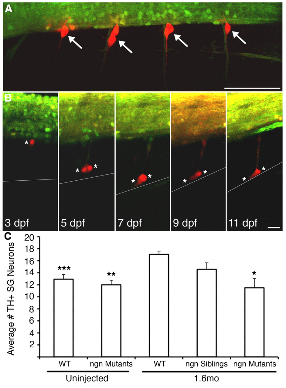
Fig. 3 Migratory DRG migrate as far ventrally as the location of the sympathetic ganglia. (A) Transgenic expression of photoconvertible Kaede in DRG allows identification of progeny and migratory paths. At 60 hpf, DRG (white arrows) in four hemisegments of a nav1.6 morphant Tg(elavl3:Kaede) embryo were selectively illuminated with UV light, as evidenced by photoconversion of Kaede from a green fluorescent molecule to red in DRG, but not nearby spinal cord, neurons. (B) At 3 dpf, a single DRG neuron (white asterisk) shows a stereotypic location at the ventral boundary of the spinal cord. At 5 dpf, the cell divides into two cells (white asterisks), but retains its axon, then migrates ventrally. At 7 dpf, both cells approach the ventral boundary of the notochord (grey line), the location of the sympathetic ganglia. The two migratory DRG cells remain in the region of the sympathetic ganglia at subsequent times (e.g. 11 dpf). (C) Consistent with migration of DRG neurons to the location of the sympathetic ganglia, the number of TH+ neurons in the sympathetic ganglia at 11 dpf is significantly greater in 1.6 morphant embryos compared with wild type (***P<0.001 versus 1.6mo wild type, nonparametric ANOVA). However, injection of 1.6mo does not produce an increase in TH+ neurons in the sympathetic ganglia in neurogenin mutants, which lack DRG neurons (**P<0.01 versus 1.6mo wild type; *P<0.05 versus 1.6mo wild type; Kruskal-Wallis nonparametric ANOVA. Scale bars: 30 μm in A; 15 μm in B.