
|
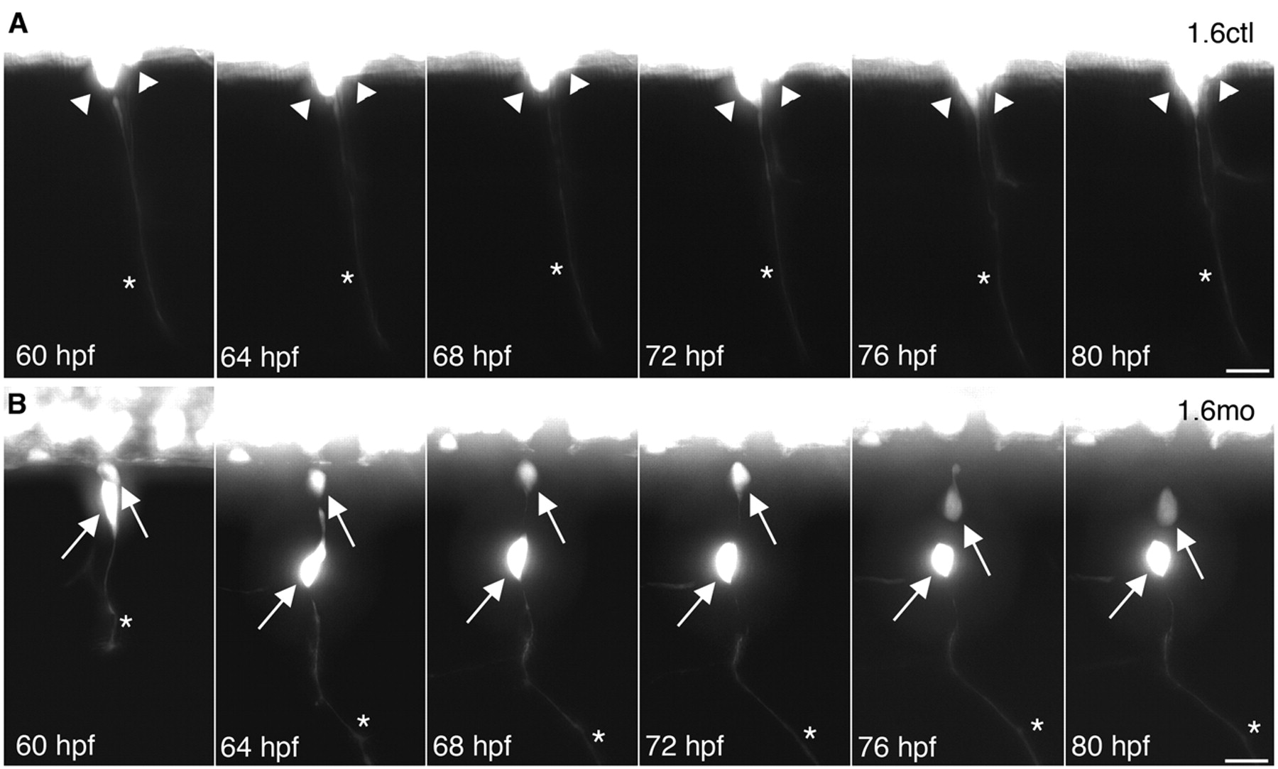
Fig. 2 DRG neurons migrate to abnormal ventral positions after they have extended axons. (A) Two adjacent DRG neurons (arrowheads) in a Tg(–3.4neurog1:GFP) control embryo extended axons (asterisk) by 60 hpf. Between 60 and 80 hpf, control DRG neurons maintain their position and remain lateral to the ventral spinal cord. Data are representative of 16 control DRG that were followed by time-lapse imaging. (B) In a 60 hpf Tg(–3.4neurog1:GFP) morphant embryo, DRG neurons (arrows) reside lateral to the ventral spinal cord and have extended axons (asterisks), similar to the control shown in A. However, at 64 hpf, one DRG neuron has migrated ventrally. By 76 hpf, the second DRG neuron has also migrated ventrally. Data are representative of 18 ventrally migrating morphant DRG neurons. Scale bars: 20 μm.