
|
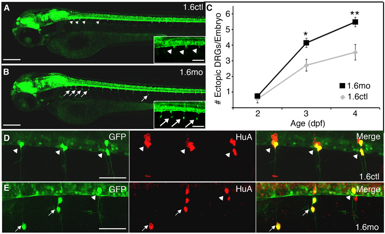
Fig. 1 DRG neurons reside in abnormal positions in nav1.6 morphants. (A) GFP+ DRG neurons show a stereotypic pattern and localize to the ventral boundary of the spinal cord (arrowheads) in each trunk hemisegment of control 3 dpf Tg(ngn:GFP) embryos. (B) By contrast, many GFP+ DRG cells reside in more ventral locations (arrows) in 1.6 morphants. Insets in A and B show segments 2-5 at higher magnification. (C) In both controls (gray diamonds) and morphants (black squares) the average number of ectopic DRG neurons per embryo increases between 2 and 4 dpf. Additionally, a larger number of ectopic DRG neurons are found in morphants than controls at 3 and 4 dpf (*P<0.01 versus 1.6mo; **P<0.001 versus 1.6mo; Kruskal-Wallis nonparametric ANOVA). (D) In 4 dpf control embryos, DRG neurons (arrowheads) express both GFP (green) and HuA (red), a marker of neuronal differentiation. (E) Ectopic DRG neurons (arrows) are also positive for both GFP and the neural differentiation marker, HuA. Embryos are mounted laterally, with anterior towards the left and dorsal at the top for all figures. Scale bars: 200 μm in A,B (70 μm in insets); 50 μm in D,E.