
|
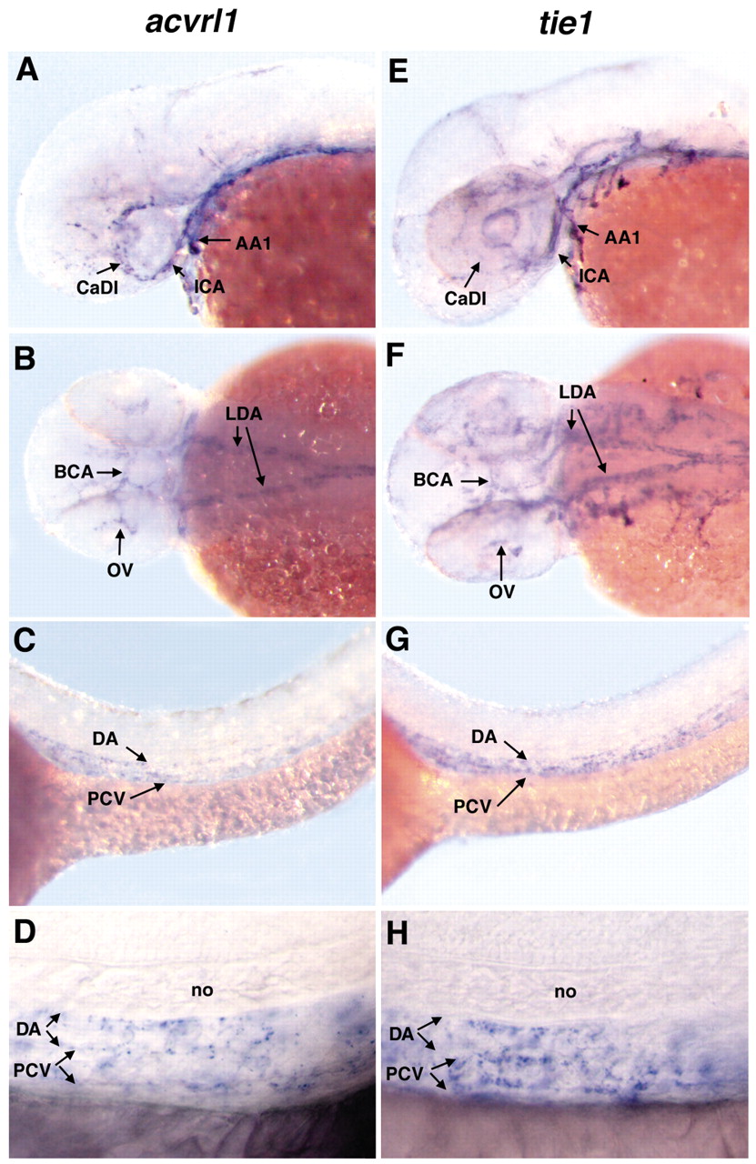
Fig. 6 Zebrafish acvrl1 is expressed predominantly in the cranial blood vessels that become dilated in vbg mutants. Whole-mount in situ hybridization using acvrl1 (A-D) or tie1 (E-H) riboprobes on 40 hpf embryos. (A,B) Expression of acvrl1 is strongest in cranial vessels, including the first arch artery (AA1), internal carotid artery (ICA), caudal division of the internal carotid artery (CaDI), basal communicating artery (BCA) and optic vein (OV). Expression in the lateral dorsal aortae (LDA) is moderate. (C,D) Very weak acvrl1 expression is present in the dorsal aorta (DA) and posterior cardinal vein (PCV) (C, low magnification; D, high magnification). In general, tie1 is more widely expressed in cranial endothelium (E,F) than acvrl1, although relative expression in the CaDI is weaker than acvrl1 expression. In the LDA (F), DA and PCV (G, low magnification; H, high magnification), tie1 expression is qualitatively similar to acvrl1. (A,C,D,E,G,H) Lateral view, anterior to the left. (B,F) Dorsal view, anterior to the left. no: notochord.