
Image
|
Figure Caption
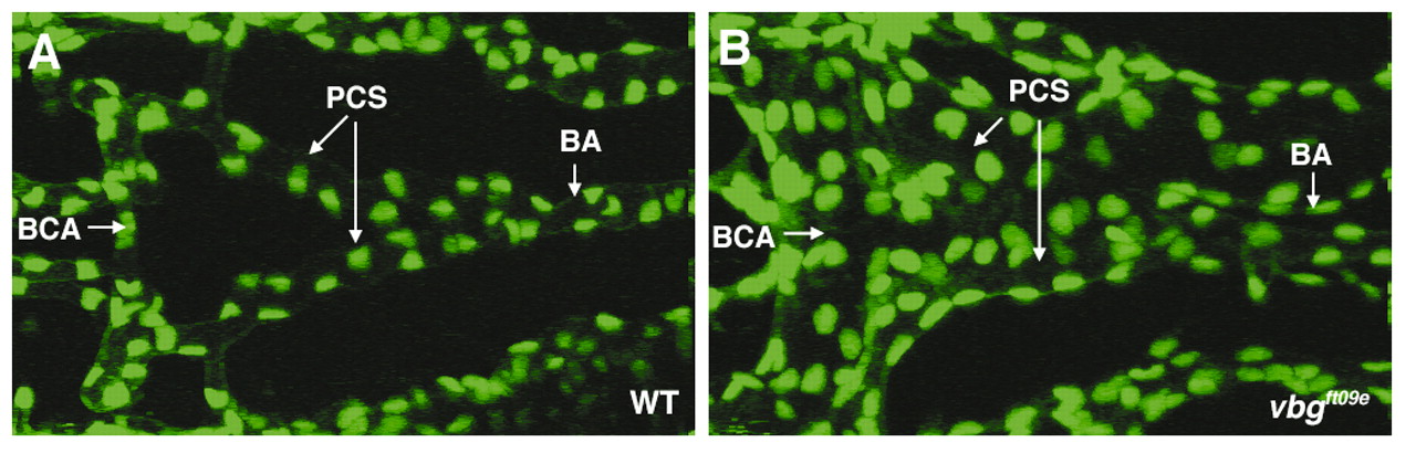
Fig. 2 Disruption of acvrl1 increases the number of endothelial cells in the basal communicating artery (BCA) and posterior connecting segments (PCS) at 2-2.25 dpf. (A,B) Representative confocal micrograph of the central cranial vasculature of (A) a vbg+;TG(fli1;nEGFP)y7 wild-type embryo, and (B) a vbgft09e;TG(fli1;nEGFP)y7 mutant embryo. The greater than two-fold increase in endothelial cell number in the BCA/PCS in vbgft09e embryos compared to wild-type embryos is significant at P<0.00001. BA, basilar artery. Dorsal views, anterior to the left.
Figure Data
Acknowledgments
This image is the copyrighted work of the attributed author or publisher, and
ZFIN has permission only to display this image to its users.
Additional permissions should be obtained from the applicable author or publisher of the image.
Full text @ Development