
|
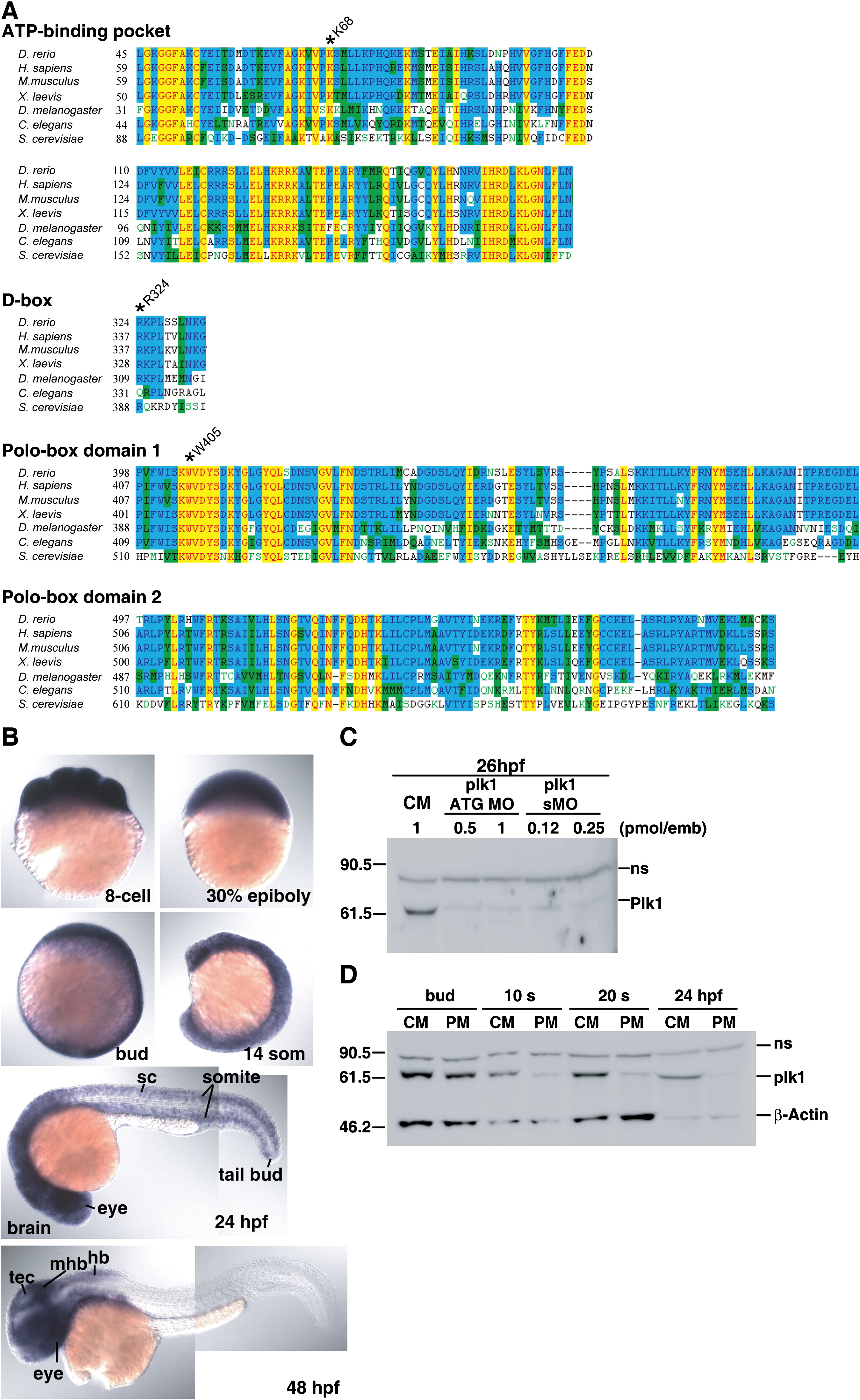
Fig. 1 Expression and morpholino-induced knockdown of Plk1 in zebrafish embryos. (A) Alignment of the ATP-binding pockets, D-boxes, and polo-box domains of Plk1 sequences from zebrafish and other species. Amino acid sequences were aligned using Clustal W. Conserved amino acids are highlighted in yellow. Asterisks indicate the mutated residues used in Fig. 7. (B) Analysis of plk1 expression by in situ hybridization in wild-type zebrafish embryos at various developmental stages: hb, hindbrain; mhb, midbrain-hindbrain boundary; sc, spinal cord; tec, tectum. (C) Embryos were injected with the indicated amounts of control MO, plk1 ATG MO, or plk1 sMO in the yolk. Whole embryo lysates were prepared at 26 hpf and subjected to western blot analysis (WB) with anti-human Plk1 antibody. The slower migrating band is a non-specific band (ns). (D) Embryos were injected with 0.5 pmol of control (CM) or plk1 ATG MO (PM), and the whole embryo lysates were subjected to WB with anti-Plk1 antibodies. The same blot was reprobed with anti-β-actin antibodies for a loading control.
Reprinted from Developmental Biology, 345(1), Jeong, K., Jeong, J.Y., Lee, H.O., Choi, E., and Lee, H., Inhibition of Plk1 induces mitotic infidelity and embryonic growth defects in developing zebrafish embryos, 34-48, Copyright (2010) with permission from Elsevier. Full text @ Dev. Biol.