
|
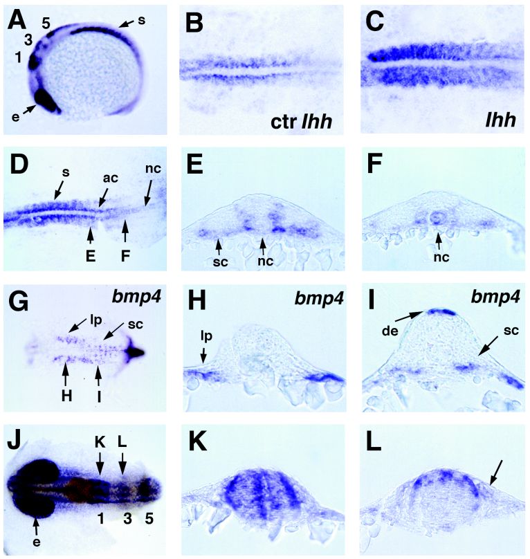
Fig. 4 Spatial expression pattern of zebrafish smad1 during somitogenesis (A–E, J–L) in comparison to eng (J) and bmp4 (G–I), and the effect of exogenous hedgehog on somitic smad1 expression (B, C). A:smad1 at the 15-somite stage, lateral view, anterior left; smad1 shows prominent expression in somites (s), eye vesicles (e), and in dorsal regions of rhombomeres 1, 3, and 5. B, C: Somitic smad1 expression is positively regulated by hedgehog signals. Fifteen-somite stage, dorsal view on trunk of flat-mounted embryos. B: Uninjected control. C: After injection of mouse Ihh mRNA (100 pg/embryo), displaying elevated smad1 mRNA levels in all regions of the somites. Similar results were obtained after injecting mRNA encoding a dominant negative version of the regulatory subunit of protein kinase A (50 pg/embryo). D:smad1 at 15-somite stage, dorsal view on trunk of flat-mounted embryo; longer stained than embryos in (B, C). The positions of transverse sections shown in E, F: are indicated. smad1 is broadly expressed in anterior somites (s), in adaxial cells (ac) of newly formed posterior somites, and in posteriormost regions of the notochord (nc). E, F: smad1, transverse sections of 15-somite-stage embryo. E: At level of the 12th somite (anterior); smad1 is strongly expressed in presumptive sclerotome (sc) in ventral regions and in medial regions of somites adjacent to the neural tube and notochord, whereas lateral and interior somitic regions and notochord are devoid of smad1 transcripts. F: At level of the 15th somite (posterior); smad1 is expressed in the notochord, whereas somitic expression is weak and largely restricted to adaxial regions. G:bmp4, 15-somite-stage, dorsal view on trunk, anterior left, head region removed. The positions of transverse sections shown in H, I: are indicated. H, I: bmp4, transverse sections of 15-somite-stage embryo. H: At level of fourth somite (anterior); bmp4 is expressed in lateral plate mesoderm (lp). I: At level of eighth somite (posterior); bmp4 is expressed in dorsal ectoderm (de) and a ventral domain of somites, most likely the presumptive sclerotome (sc). J:smad1 (blue) and Eng (brown, detected by immunostaining) at 15-somite-stage, dorsal view on head region. eng expression marks the region of the midbrain–hindbrain boundary. Rhombomere numbers are indicated. The positions of transverse sections shown in (K) and (L) are indicated with arrows. K, L:smad1, transverse sections of 15-somite-stage embryo at level of first (K) and third (L) rhombomere. First rhombomere displays smad1 expression in three dorsoventral stripes, a medial stripe, and two lateral stripes at the periphery of the brain. At the level of the third rhomobomere, smad1 displays strongest expression in dorsal regions of the brain. In addition, smad1 is expressed in dorsal cells outside of the neural tube (indicated by an arrow in L), which might represent migrating neural crest cells. Abbreviations: ac, adaxial cells; de, dorsal ectoderm; e, eye vesicles; lp, lateral plate; nc, notochord; s, somites; sc, sclerotome.