
|
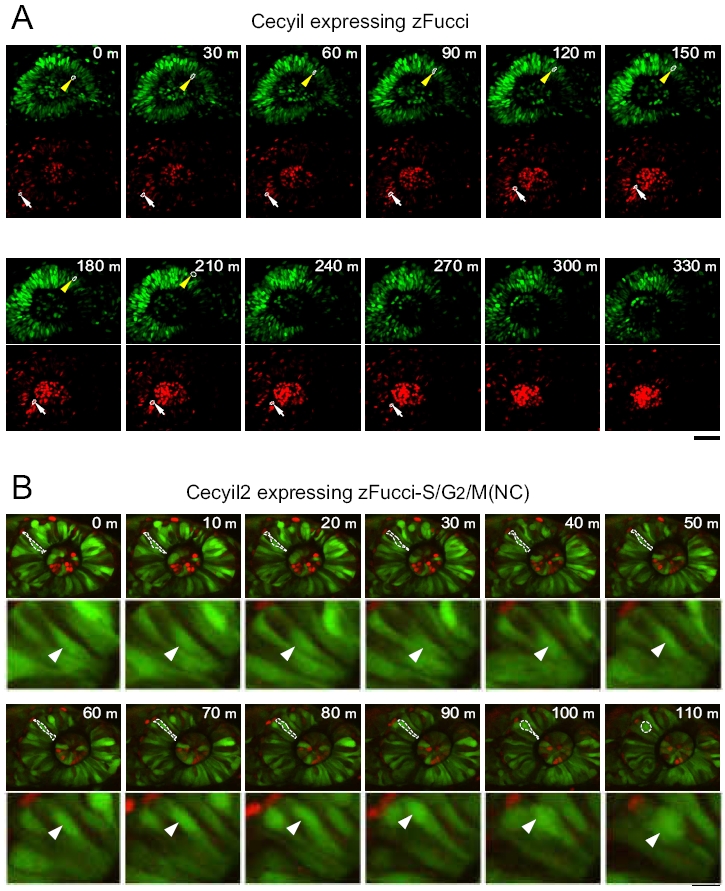
Fig. S4 Interkinetic nuclear migration in the retina at early stages. An embryo at 22 hpf was anesthetized with 0.00168% Tricaine and embedded in 0.3% agar on a culture dish with the eye facing up. Fluorescence images were collected using an Olympus FV1000 upright confocal microscope to visualize interkinetic nuclear migration, namely, the apical-basal movement of nuclei in phase with the cell cycle. The developing lens at the center of the eye showed intense orange signal, suggesting early differentiation of the lens. (A) Fluorescence images of an eye from a Cecyil embryo expressing zFucci: mAG-zGem(1/100) (Top) and mKO2-zCdt1(1/190) (Bottom). Each nucleus in the sheet emitted either green or orange fluorescence. (Scale bar, 50 μm.) (B) Fluorescence images of an eye from a Cecyil2 embryo expressing zFucci-S/G2/M(NC): mAG-hGem(1/60) and mKO2-zCdt1(1/190) (merged). (Scale bar, 50 μm.)