
|
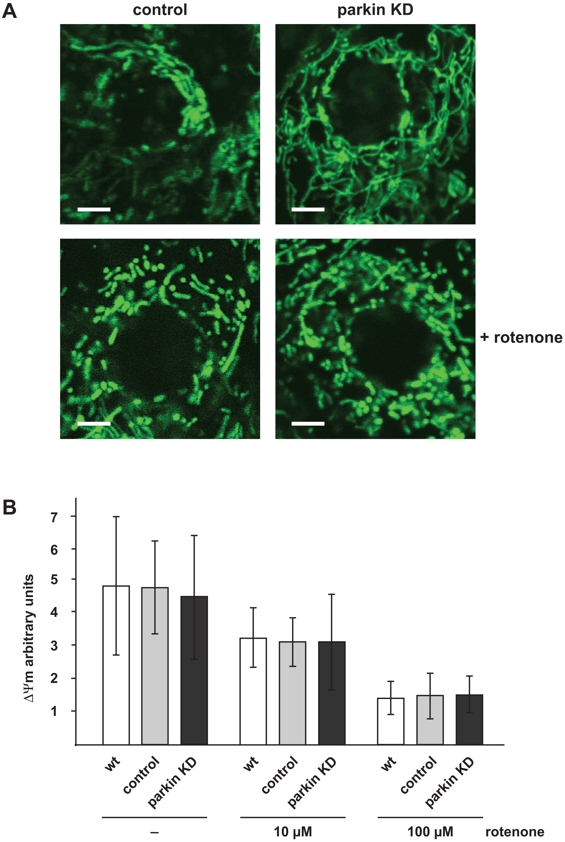
Fig. 9 Mitochondrial morphology and membrane potential is not affected in parkin-deficient zebrafish.
(A) Mitochondrial morphology in not altered in parkin knockdown zebrafish embryos. Live cell imaging of mitochondria in one-day-old parkin-deficient zebrafish and control-injected zebrafish. Mitochondria were visualized by the transient expression of GFP targeted to mitochondria (green). A transient knockdown of parkin in zebrafish did not cause mitochondrial fragmentation in the outer skin of zebrafish embryos (upper images). Mitochondrial fragmentation induced by rotenone (100 μg/l, 6 h) was not aggravated in parkin-deficient zebrafish embryos (lower images). Scale bar 5 μm. (B) The membrane potential of total mitochondria isolated from parkin-deficient zebrafish embryos is not altered in comparison to mitochondria isolated from wildtype or control-injected embryos, neither under basal conditions nor after rotenone treatment. Isolated mitochondria from one-day-old wildtype, control-injected and parkin knockdown zebrafish were either left untreated or stressed with different concentrations of rotenone and subsequently incubated with the fluorescent dye JC-1. The mitochondrial membrane potential is represented as the ratio of intra-mitochondrial JC-1 (orange) and cytosolic JC-1 (green). Quantification is based on three independent experiments. KD: knockdown.