
|
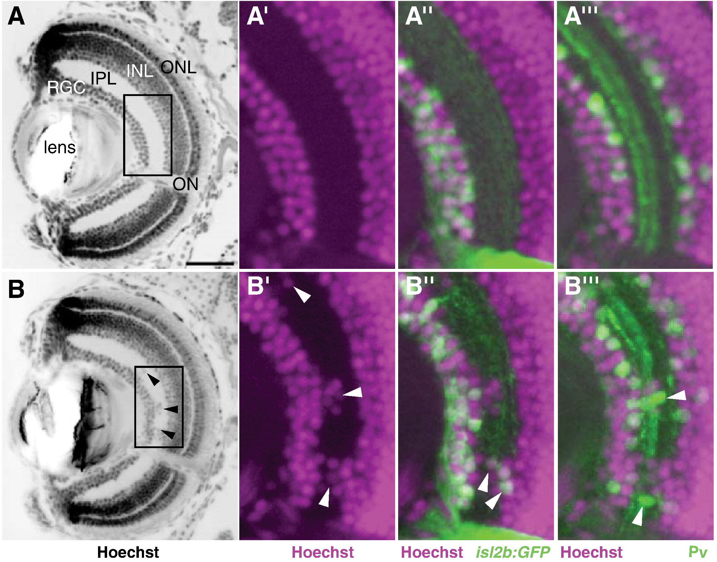
Fig. 6 Retinal lamination is disrupted in nev. Coronal sections through a WT eye (A–A″′) and a nev eye (B–B″′) at 5dpf. (A and B) Hoechst 33342 stain to visualize lamination. The retinal ganglion cell (RGC) layer, inner plexiform layer (IPL), inner nuclear layer (INL), and outer nuclear layer (ONL) are all clearly visible at this stage. Arrowheads in B show displaced cells in the IPL of nev. Boxed regions are magnified in A′–A″′ and B′–B″′ and show nuclei labeled with Hoechst 33342 (magenta; A′–A″′, B′–B″′), RGCs labeled with isl2b:GFP (green; A″, B″), and a subset of amacrine cells labeled with anti-parvalbumin (green; A″′, B″′). In nev, RGCs and amacrine cells are intermingled in the IPL. Arrowheads in B″ and B″′ show displaced RGCs and amacrine cells in the IPL, respectively. ON, optic nerve. Scale bar = 50 μm.
Reprinted from Developmental Biology, 344(2), Pittman, A.J., Gaynes, J.A., and Chien, C.B., nev (cyfip2) Is required for retinal lamination and axon guidance in the zebrafish retinotectal system, 784-794, Copyright (2010) with permission from Elsevier. Full text @ Dev. Biol.