
|
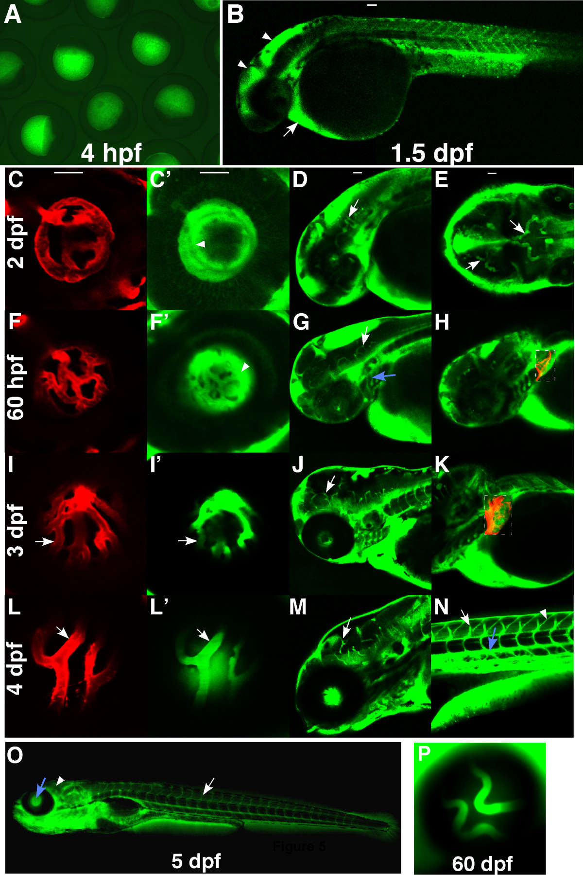
Fig. 5 Expression pattern of DBP-EGFP in the blood plasma of l-fabp:DBP-EGFP transgenic zebrafish. Images were obtained from Tg(l-fabp:DBP-EGFP;flk1:mCherry) fish to visualize DBP-EGFP (green) and endothelial cells lining blood vessels (red)(mCherry). The fish were oriented with anterior to the left. All panels are side views except E. (A) green fluorescence in eggs laid by female Tg(l-fabp:DBP-EGFP) fish. (B) DBP-EGFP expression at 1.5 dpf. (C, C′, D & E) DBP-EGFP expression at 2 dpf. The central arteries (arrows) are the first distinguishable blood vessels. (F, F′, G & H) DBP-EGFP expression at 60 hpf. The dashed rectangles in H and K are merged pictures of red and green channels. The branchial arches (blue arrow) can be differentiated at the time, but boundaries of HV are still not clear (arrowhead). At 3 dpf (I, I′, J & K), the boundaries of the HV become sharp (arrows in I&I′). More vessels appear in the liver (dashed rectangle) and more brain vessels can be seen (arrows in G and J). At 4 dpf (L, L′, M & N), the EGFP-infused hyaloid and brain vessels can be easily differentiated from the fluorescent background (arrows in L, L′ and M), as can the intersegmental vessels (arrow in N) and the dorsal aorta (blue arrow). Accumulation of DBP-EGFP in the myotomal boundaries (arrowhead) (N) is observed from leakage out of the vasculature. At 5 dpf (O), the HV (blue arrow) and the trunk vessels (arrow) are distinguishable, but the brain vessels are not (arrowhead), due to the increased fluorescent background in the brain. (P) At 60 dpf, the HV is still distinguishable.