
Image
|
Figure Caption
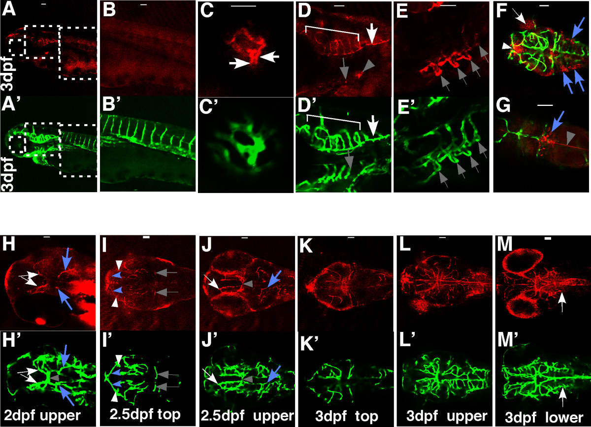
Fig. 2 Spatial and temporal expression of claudin-5 in the developing BBB. Tg(flk1:EGFP) embryos at 2 dpf, 2.5 dpf, and 3 dpf were stained with a monoclonal claudin-5 antibody. Confocal images of whole mount embryos were analyzed for claudin-5 expression (red) (A-E and H-M; Alexa Fluor 568 labeled secondary antibody) in developing blood vessels (green) (A′-E′ and H′-M′; EGFP labeled vascular endothelial cells; F&G, merged pictures). All the samples are oriented with anterior to the left. A-E and A′-E′, lateral views; other panels, dorsal views. Scale bars: 50 μm.
Figure Data
Acknowledgments
This image is the copyrighted work of the attributed author or publisher, and
ZFIN has permission only to display this image to its users.
Additional permissions should be obtained from the applicable author or publisher of the image.
Full text @ BMC Dev. Biol.