
|
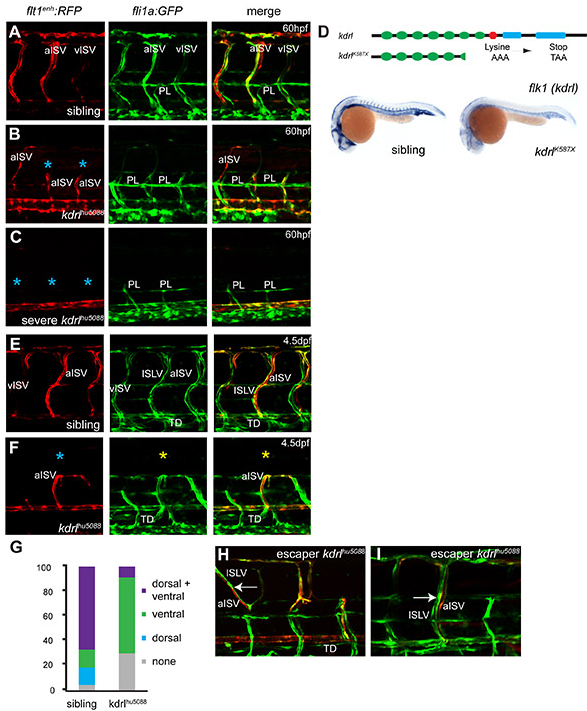
Fig. S3 Arteries are required for lymphatic development. (A-C) 60 hpf sibling and mutant kdrlhu5088 in fli1a:GFP; flt1enh:RFP double transgenic embryos reveal that PL formation is normal, whereas the primary sprouts can be affected differently (indicated by the yellow asterisk) in mutants. (D) Kdrlhu5088 failed to complement kdrlt21588 (Habeck et al., 2002) and sequencing revealed an A-to-T transversion, changing a lysine at amino acid position 587 into a premature stop (K587X) resulting in reduced kdrl expression. (E,F) Lymphatic vasculature establishment in 4.5 dpf sibling and mutant kdrlhu5088 fli1:GFP; flt1enh:RFP double transgenic embryos with normal or with reduced primary sprouts at 4.5 dpf. Mutants have reduced primary sprouting (indicated by blue asterisks) and lack dorsal migration of LECs (indicated by yellow asterisks), whereas ventral migration is still present (G). Quantification of dorsal and ventral migration along arteries. Mutants with thoracic duct (n=5, total of 23 arteries) without dorsal migration, whereas the ventral migration along these arteries is mostly retained. (H,I) kdrlhu5088 escapers in fli1:GFP, flt1enh:RFP double transgenic embryos at 4.5 dpf. LECs (arrows) migrate along escaping arteries that did succeed in growing dorsally.