
|
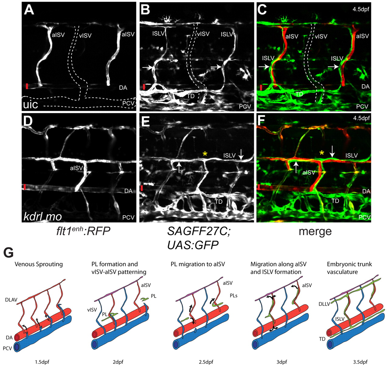
Fig. 4 Intersegmental arteries are required for LEC migration. (A-C) flt1enh:RFP (A), SAGFF27C;UAS:GFP (B) and merged (C) unilateral confocal images of control embryos at 4.5 dpf showing lymphatic patterning along aISVs in normal blood vasculature. Arrows indicate ISLVs. (D-F) flt1enh:RFP (D), SAGFF27C;UAS:GFP (E) and merged (F) unilateral confocal images of kdrl morpholino injected embryos at 4.5 dpf showing LEC migration along arteries that cross-connect abnormally at the horizontal myoseptum, and that do not extend into the dorsal half (indicated by yellow asterisk) of the respective somite. Arrows indicate LECs along the surface of an abnormal aISV. Vertical red bars indicate the DA. (G) Model of lymphatic patterning within the zebrafish trunk. Secondary sprouts from the PCV that do not connect to primary intersegmental vessels migrate to the horizontal myoseptum region where they constitute a pool of PLs. PLs will migrate ventrally or dorsally to form the thoracic duct and ISLVs, respectively, and use arteries as their substrate.