
|
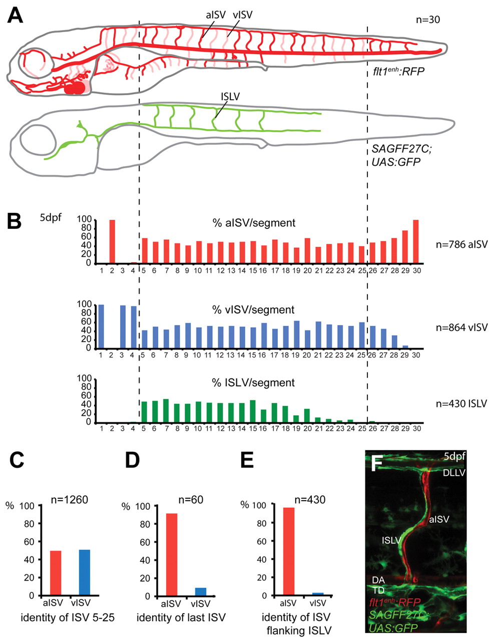
Fig. 2 Arteriovenous and lymphatic patterning in the zebrafish trunk. (A) Schematic representation of arteries, veins and lymphatic vessels at 5 dpf, based on expression of flt1enh:RFP and SAGFF27C;UAS:GFP over 25-30 segments in 30 embryos (segment numbers vary in zebrafish embryos, with an average of 27.2 segments or 54.4 segmental positions per embryo). (B) Quantification of vessel type distribution per segment, indicating aISV in red bars, vISVs in blue bars and ISLVs in green bars. (C) Quantification of aISV/vISV identity in segments 5-25, n=1260 segmental positions scored. (D) Quantification of aISV/vISV identity of the caudal-most ISV/side, n=60 indicating a strong preference for aISV over vISV in the last ISV position. (E) Quantification of arterial and venous ISVs abutting an ISLV, n=430 intersegmental vessel pairs scored. (F) Overlaid confocal images of flt1enh:RFP+ aISV and SAGFF27C;UAS:GFP+ ISLV. aISV, arterial ISV; DA, dorsal aorta; DLLV, dorsal longitudinal lymphatic vessel; ISLV, intersegmental lymphatic vessel; TD, thoracic duct; vISV, venous ISV.