
Image
|
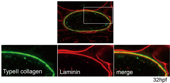
Figure Caption
Fig. S8 The medial layer appears to consist of type II collagen. Type II collagen was detected external to the laminin-rich inner layer. The boxed area is enlarged in the lower panels. Green, type II collagen; red, laminin; yellow, merged expression of type II collagen and laminin. Transverse sections through the trunk region of an embryo at 32 hpf are shown.
Acknowledgments
This image is the copyrighted work of the attributed author or publisher, and
ZFIN has permission only to display this image to its users.
Additional permissions should be obtained from the applicable author or publisher of the image.
Full text @ Development