
|
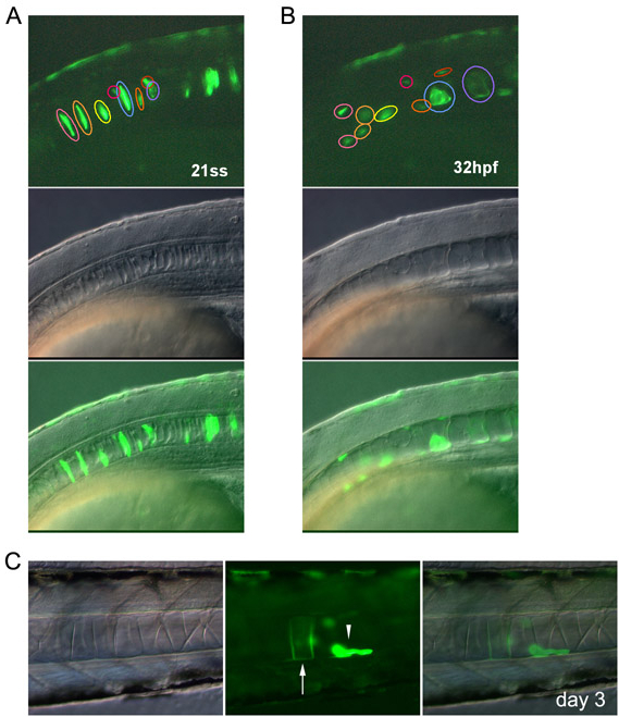
Fig. S5 Intercalated notochord cells develop into vacuolated and non-vacuolated epithelial cells. (A-C) Notochord cells were tracked by GFP expression under the promoter of the floating head gene (Flh-GFP). (A,B) Time-lapse images of the notochord cells of an embryo at 21 ss and 32 hpf. Cells indicated by pink and yellow circles developed into non-vacuolated cells. The cell indicated by a blue circle developed into a vacuolated cell. (C) A non-vacuolated epithelial cell observed on day 3. Vacuolated and non-vacuolated cells are indicated by the arrow and arrowhead, respectively. Images are lateral views with anterior to the left.