
Image
|
Figure Caption
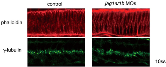
Fig. S3 Normal cell morphology of the notochord is established in the jagged 1 knockdown embryos at 10 ss. Embryos were stained by Rhodamine-phalloidin or anti γ-tubulin. Normal intercalated cell morphology of the notochord was observed with phalloidin staining. Notochord cell polarities were well established, as assessed by the central localization (basal) of γ-tubulin in the notochord in the jag1a/1b-knockdown embryos (jag1a/1b MOs). Side views of the notochord cells in 10 ss embryos are shown.
Acknowledgments
This image is the copyrighted work of the attributed author or publisher, and
ZFIN has permission only to display this image to its users.
Additional permissions should be obtained from the applicable author or publisher of the image.
Full text @ Development