
|
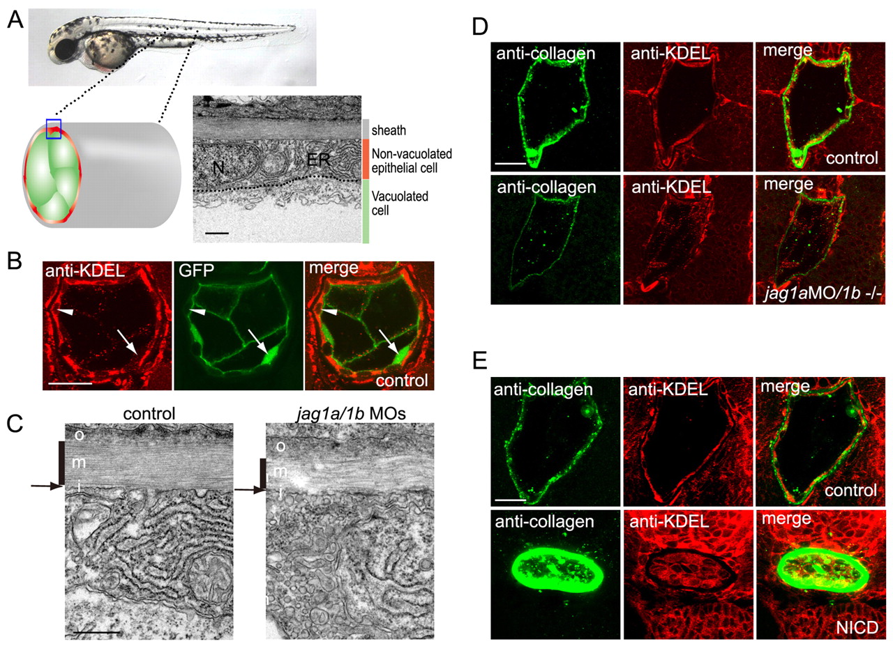
Fig. 5 Jagged 1a/1b is required for notochord extracellular matrix sheath formation. (A) Non-vacuolated epithelial cells contained abundant rough ER compared with vacuolated cells. N, nuclei; ER, endoplasmic reticulum. Transmission electron micrographs (TEMs) of transverse sections of a wild-type embryo on day 2. (B) Non-vacuolated cells were rich in rough ER. ER was stained by an anti-KDEL antibody (red), and vacuolated cells were revealed by the 214A-GFP transgenic line (green). Arrowheads indicate anti-KDEL-positive non-vacuolated cells. Arrows indicate GFP-positive vacuolated cells. (C) The medial layer of the peri-notochordal basement membrane was thinner in the jag1a/1b knockdown embryos than in wild-type embryos. TEM of transverse sections. o, outer; m, medial; i, inner. Black vertical lines indicate the medial layer of the sheath. Arrows indicate the thin inner layer. (D) jag1a/1b knockdown resulted in reduced type II collagen deposition and fewer cells with abundant rough ER in the notochord. (E) Notch activation increased type II collagen deposition and ER-positive cells in the notochord. Control siblings (control) or double transgenic Tg(UAS:myc-Notch1a-intra);Tg(hsp70:Gal4) embryos (NICD) were heat shocked at 3 ss. A-E are transverse sections of day 2 embryos. Scale bars: 0.5 μm in A,C; 20 μm in B,D,E.