
|
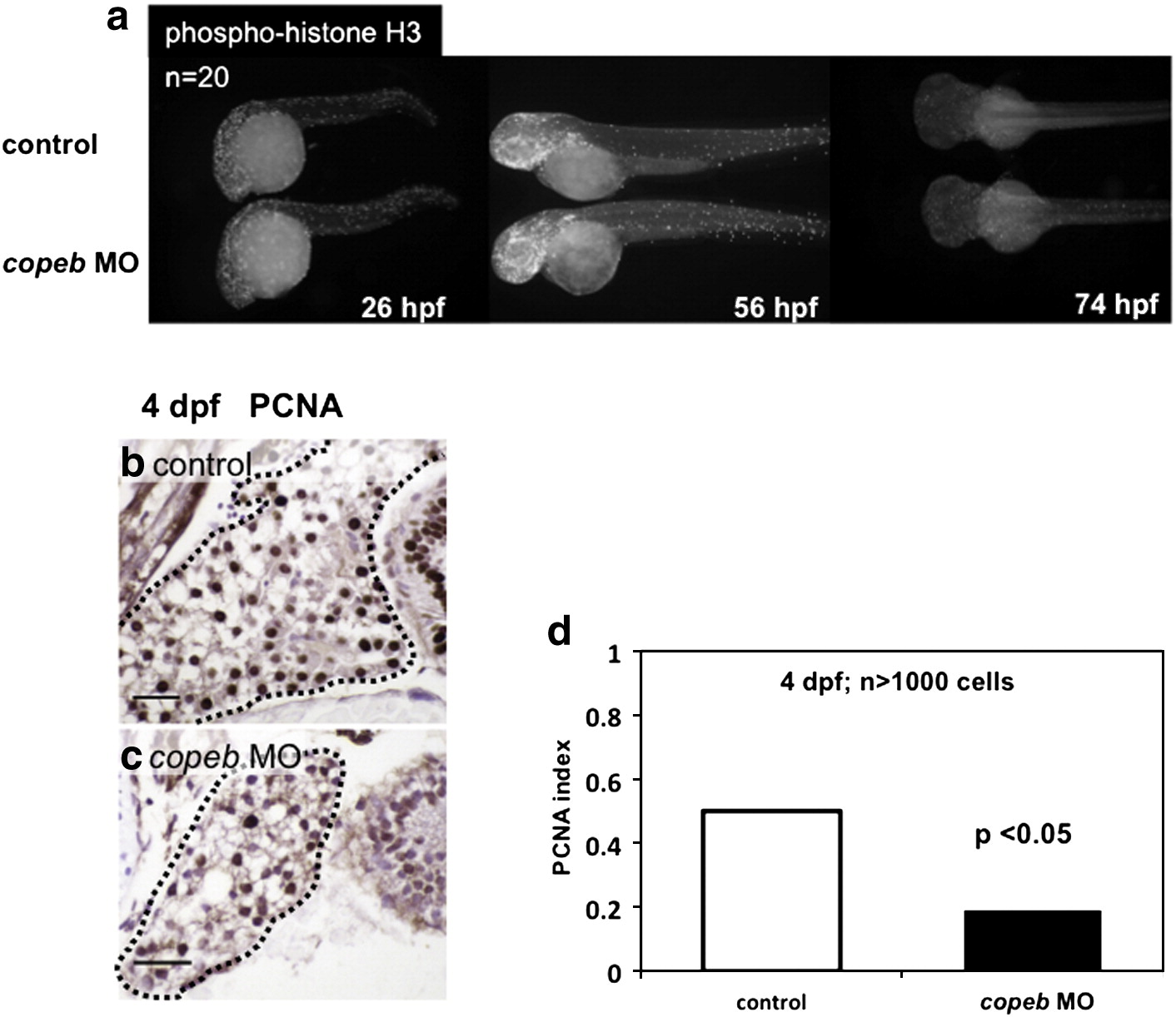
Fig. 5 Decreased endoderm-derived organ size in copeb morphants is due to decreased proliferation. (a) Proliferation in whole embryos was examined at 26, 56, and 74 hpf by immunofluorescence using an antibody against phosphorylated Histone H3 (p-histone H3) to identify cells in the G2/M phase of the cell cycle. High levels of proliferation are found in the head, eyes and tails of both morphants and controls; n = 20 for each sample at each time point. (b–d) Hepatocyte proliferation was assessed by nuclear PCNA staining of serial sections of the liver (outlined) in control embryos (b) and copeb morphants (c) on 4 dpf and was quantified in (d). scale bar = 20 microns. At least 1000 hepatocytes in at least 2 control and 3 morphant embryos were scored for each sample.
Reprinted from Developmental Biology, 344(1), Zhao, X., Monson, C., Gao, C., Gouon-Evans, V., Matsumoto, N., Sadler, K.C., and Friedman, S.L., Klf6/copeb is required for hepatic outgrowth in zebrafish and for hepatocyte specification in mouse ES cells, 79-93, Copyright (2010) with permission from Elsevier. Full text @ Dev. Biol.