
|
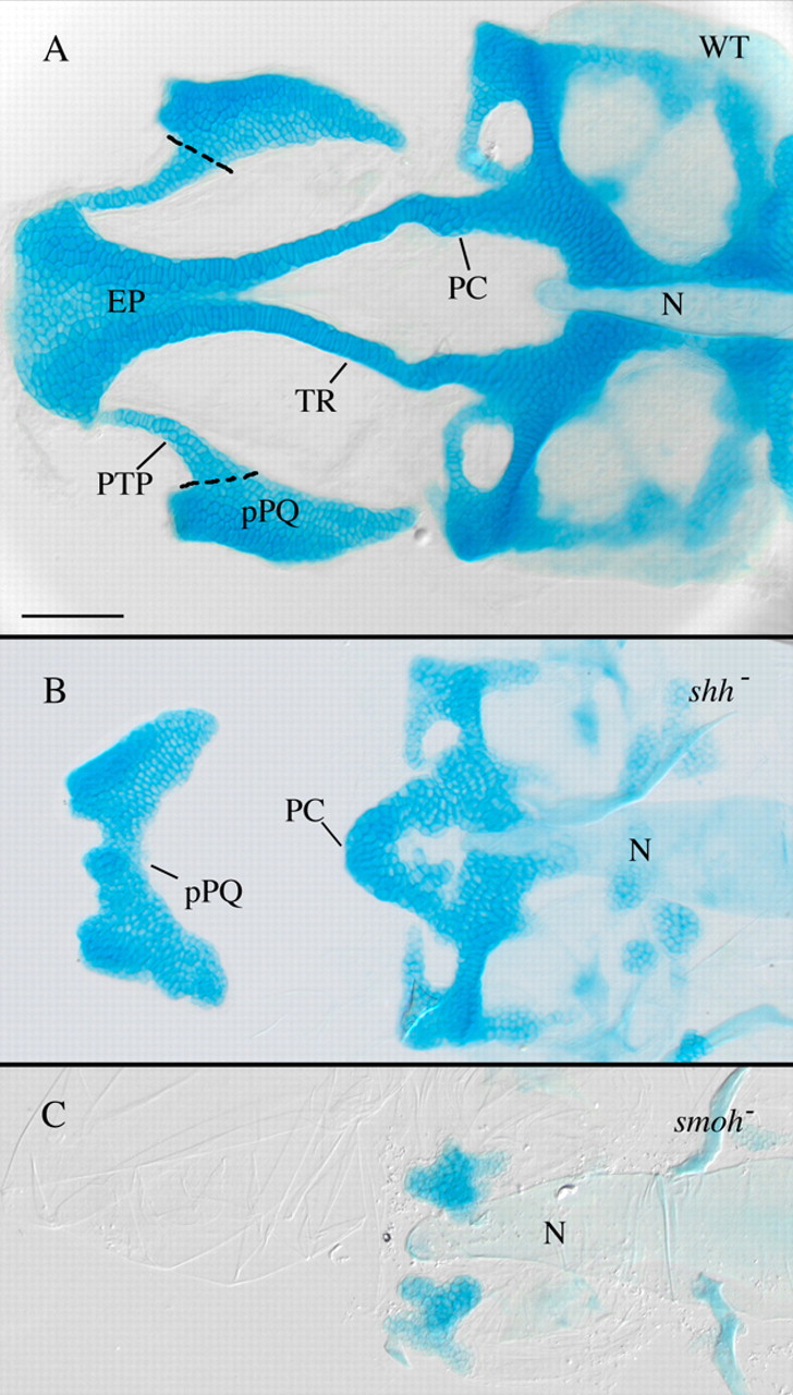
Fig. 1 Anterior craniofacial cartilages are especially sensitive to loss of Hh signaling. (A) Flat-mounted 4 dpf wild-type anterior craniofacial cartilages. The anterior neurocranium consists of the trabeculae (TR) and the ethmoid plate (EP). The polar cartilages (PC) fuse with TR joining the anterior and more posterior neurocranium. The dorsal first arch cartilage palatoquadrate (pPQ) and its pterygoid process (PTP) are flat mounted with the neurocranium. (B) Hypomorphic shh- embryos have variable anterior neurocranium defects, including loss of anterior neurocranium and PTP with fusion of pPQ across the midline. (C) Loss of Hh signaling in smo- embryos eliminates most neurocranium cartilages obscuring analysis of defects specific to the anterior neurocranium. N, notochord; WT, wild type. Dorsal views, anterior is leftwards in all panels. Scale bar: 50 μm.