
Image
|
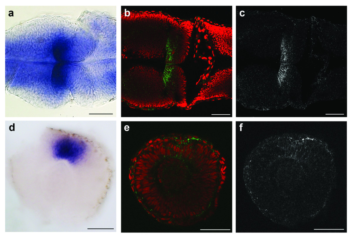
Figure Caption
Fig. 4 Recombinant monoclonal antibodies recapitulate their gene expression patterns on wholemount fixed zebrafish embryos. (a-c) Dorsal views of a 24 hpf zebrafish embryo showing flrt3 gene expression by in situ hybridization on the midbrain side of the mid-hindbrain boundary (a) and antibody staining with SI3-Flrt3 [green in (b), white in (c)]. (d-f) Lateral view of a dissected 24 hpf eye showing expression of the unc5b gene in the dorsal retina (d) and antibody staining with SI2-Unc5b [green in (e), white in (f)]. (b, e) red = DAPI nuclear stain. Scale bars = 50 μm.
Acknowledgments
This image is the copyrighted work of the attributed author or publisher, and
ZFIN has permission only to display this image to its users.
Additional permissions should be obtained from the applicable author or publisher of the image.
Open Access.
Full text @ BMC Biol.