
Image
|
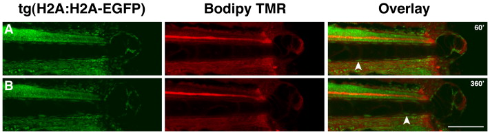
Figure Caption
Fig. 2 Cells from the notochord contribute to the regeneration blastema. Strip of images taken from two-color time-lapse of a transgenic tg(H2A:H2A-EGFP) larvae injected with Bodipy-TMR, with the time of image capture shown in minutes after amputation. Tracking of individual cells during this period of time indicates that cells from the notochord (white arrowheads) are rapidly deposited into the blastema (see also Movie S1). Scale bar 50 μm.
Acknowledgments
This image is the copyrighted work of the attributed author or publisher, and
ZFIN has permission only to display this image to its users.
Additional permissions should be obtained from the applicable author or publisher of the image.
Reprinted from Developmental Biology, 327(1), Rojas-Muñoz, A., Rajadhyksha, S., Gilmour, D., van Bebber, F., Antos, C., Rodríguez Esteban, C., Nüsslein-Volhard, C., and Izpisúa Belmonte, J.C., ErbB2 and ErbB3 regulate amputation-induced proliferation and migration during vertebrate regeneration, 177-190, Copyright (2009) with permission from Elsevier. Full text @ Dev. Biol.