
|
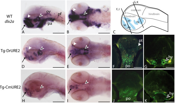
Fig. 4 Expression of URE2-GFP reporter constructs in the brain of 48 hpf zebrafish. Expression patterns obtained by in situ hybridization using a dlx2a cDNA probe in wild-type embryos (A, B) or a GFP probe in Tg-DrURE2 (D, E) and in Tg-CmURE2 embryos (H, I). Immunolocalization of GFP proteins on sectioned embryos of the Tg-DrURE2 (F, G) and Tg-CmURE2 (J, K). Expression in the telencephalon is comparable for the endogenous gene and the two transgenes (black arrow in A, B, D, E, H, I). Expression in the dorsal domain of the prethalamus (white arrowhead) in Tg-DrURE2 for gfp mRNA (D, E) and GFP proteins (F) is not observed in Tg-CmURE2 (H-J). Expression of GFP in the hypothalamus (black arrowhead) was restricted to lateral cells in Tg-CmURE2 (H, I, K) compared to Tg-DrURE2 (D, E, G). Panels A, D, H are lateral views, B, E, I are ventral views. Plan for the transversal sections presented in F-G and J-K are localized on the scheme in panel C. Blue domains in the scheme are the forebrain expression domains described for dlx genes: the telencephalic domain being the subpallium (sp, black arrow); the diencephalic domains being the preoptic area (po), prethalamus (pt, white arrowhead) and hypothalamus (hy, black arrowhead). Scale bars: A, B, D, E, H, I, 250 μm; F, G, J, K, 50 μm.