
|
Fig. 1 Loss of SDF1a Leads to a Stronger Phenotype than Loss of Cxcr4b
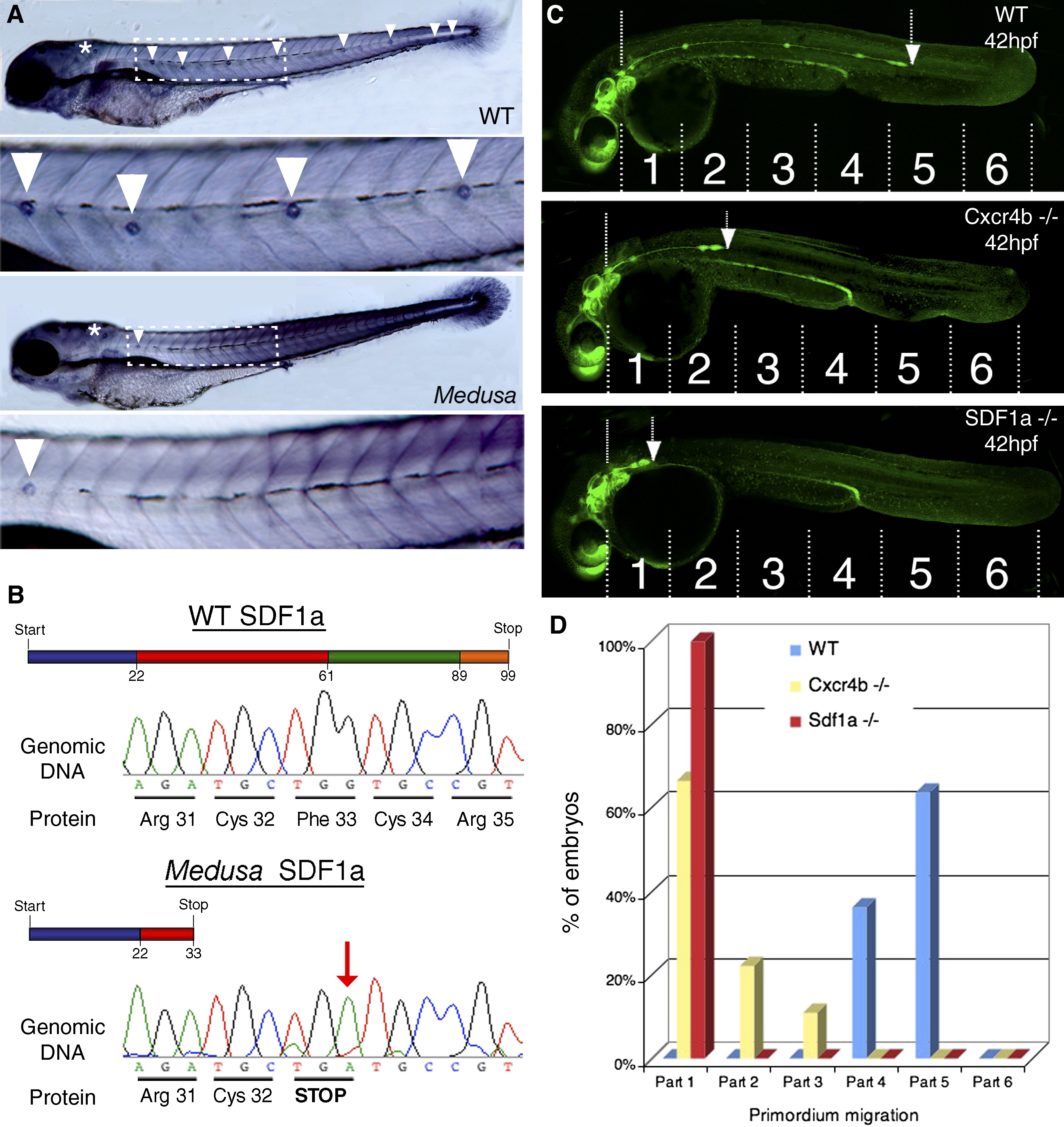
(A) Neuromast deposition was detected at 4.5 days postfertilization (dpf) by alkaline phosphatase staining. Wild-type embryos show a zebrafish-specific neuromast pattern (upper panels, overview and magnified view of dashed box). In medusa mutants, the neuromast number in the posterior lateral line is reduced (arrowheads), whereas anterior neuromasts are still present (asterisk) (lower panels, overview and magnification of dashed box).
(B) Schematic representation of a full-length SDF1a protein in the wild-type and a mutated SDF1a protein in medusa mutants (the colors represent the four exons of the sdf1a gene). A point mutation at nucleotide position 99 converts phenylalanine into a stop codon (red arrow). This nonsense mutation truncates SDF1a after 32 amino acids in medusa mutants.
(C) The lateral-line-primordium migration was analyzed in wild-type, cxcr4b, and sdf1a mutant embryos at 42 hpf. Six equal sections from the ear to the end of the tail were defined, and an equal number of embryos (n = 37) were classified according to the leading-edge position of the primordium (arrows).
(D) Quantification of results from (C) shows that SDF1a mutants have a stronger migratory defect than do cxcr4b mutants (compare red and yellow bars).