
|
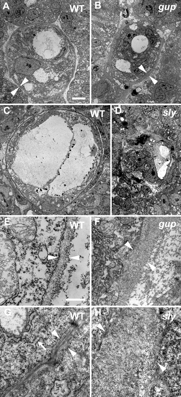
Fig. 6 Electron micrographs of wild-type, sly and gup mutant embryos. Sections through notochords of (A,E) a wild-type (WT) embryo at 28 hpf; (B,F) a gup mutant at 28 hpf; (C,G) a wild-type embryo at 32 hpf; (D,H) a sly mutant at 32 hpf. Notochordal sheath (between arrowheads) of WT (A,E) and gup (B,F) 28 hpf embryos are shown with intracellular space to the left and extracellular to the right. Wild-type basement membrane (E) appears as three distinct layers; closest to the cell membrane is a faint diffuse layer, which probably contains laminin, followed by a fibrous layer running parallel to the plane of section and finally an outermost fibrous layer running perpendicular to the plane of section, both of which are likely to be collagen rich. Conversely, in a gup mutant sibling (F), a thick disorganized layer is observed. Similarly, in the more mature notochordal sheath of a 32 hpf wild-type embryo (G), a well organized sheath is observed (between arrowheads). In a sly mutant sibling (H), however, the sheath is completely disorganized (between arrowheads). (G) An example of wild-type rough endoplasmic reticulum is shown (arrow). Scale bars: in A 5 μm for A-D; in E, 0.5 μm for E-H.