
|
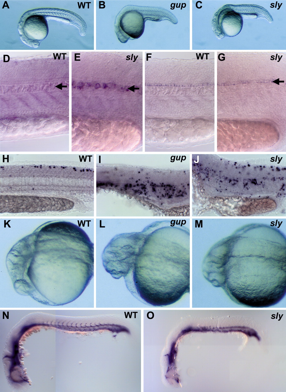
Fig. 1 Morphological and molecular analysis of sly and gup mutants. Lateral views show morphological phenotypes of wild-type (WT) (A), gup (B) and sly (C) mutant embryos at 22 hpf. (D) Whole-mount in situ hybridization shows that by 28 hpf ehh expression is no longer observed in the wild-type vacuolated notochord (arrow). (E) In sly mutants the notochord remains undifferentiated and expression of ehh is maintained (arrow). (F,G) In WT and sly mutants, the expression of twhh is identical and restricted to the floor plate (arrows). (H) TUNEL analysis at 28 hpf shows that apoptosis is restricted to the dorsal neural tube in WT embryos. (I,J) In both gup (I) and sly (J) mutant embryos, more extensive apoptosis is observed along the embryonic midline. (K-M) Close-up images of WT (K), gup (L) and sly (M) mutant embryos at 22 hpf show the small retina and protruding lens of the mutants. (N,O) Whole-mount in situ hybridization to detect expression of fli1, a marker of vascular progenitor cells, shows that the inter somitic vessels seen forming in 22 hpf WT embryos (N) fail to form in sly mutants (O).