
Image
|
Figure Caption
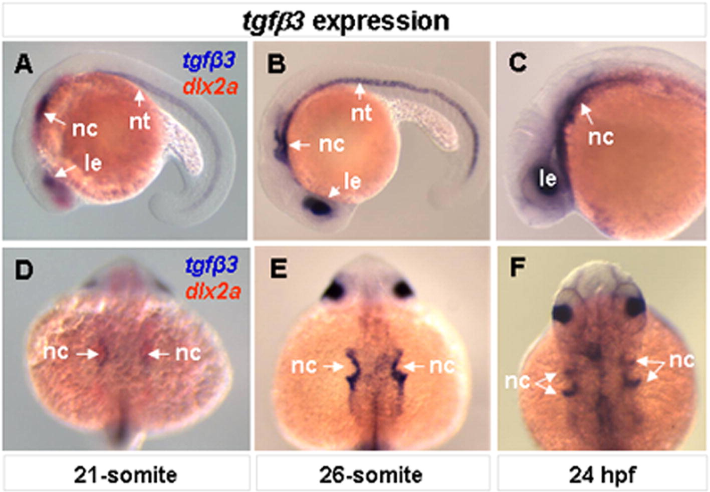
Fig. S2 Embryonic expression of tgfβ3. Whole-mount lateral (A–C) and dorsal (D–F) views are shown. At the 21-somite stage, tgfβ3 is expressed in the lens, migrating neural crest cells and notochord (A, D). Double-color in situ hybridization with known migrating neural crest marker, dlx2a, shows that tgfβ3 is expressed in a subpopulation of the migrating neural crest cells (A, D). The expression of tgfβ3 in the migrating neural crest cells intensifies in streams 2 and 3 at the 26-somrte stage (B, E) and in 24 hpf (C, F) embryos.
Figure Data
Acknowledgments
This image is the copyrighted work of the attributed author or publisher, and
ZFIN has permission only to display this image to its users.
Additional permissions should be obtained from the applicable author or publisher of the image.
Reprinted from Mechanisms of Development, 128(7-8), Cheah, F.S., Winkler, C., Jabs, E.W., and Chong, S.S., tgfbeta3 Regulation of Chondrogenesis and Osteogenesis in Zebrafish is Mediated Through Formation and Survival of a Subpopulation of the Cranial Neural Crest, 329-344, Copyright (2010) with permission from Elsevier. Full text @ Mech. Dev.