
|
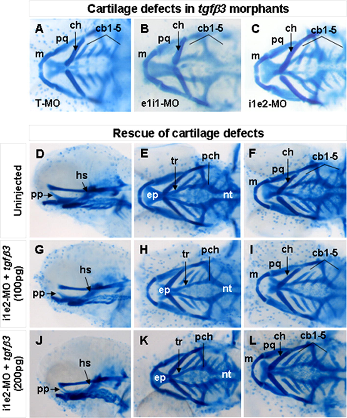
Fig. S1 Craniofacial cartilage phenotypes in 4 dpf morphents injected with different tgfβ3-targeting antisense MOs and their rescue by co-injection of tgfβ3 mRNA. Whole-mount ventral (A–C, F, I, L), lateral (D, G, J), and dorsal (E, H, K) views are shown. Similar cartilage defects are detected in hatchlings injected with translation blocking T-MO (A), splice inhibition elit-MO (B) and i1e2-MO (C). Compared with uninjected wild-type hatchlings (D–F), co-injection of 100 pg of tgfβ3 mRNA rescued the cartilage defects in 27% of the i1e2 MO-injected hatchling (G–I), whereas 33% of the i1e2 MO-injected hatchlings were rescued by co-injection of 200 pg of tgfβ3 mRNA (J–L), cbl-5. ceratobranchial arches 1–5; ch, ceratohyal; ep, ethmoid plate; hs, hyosymplectic; m, Meckel’s cartilage; nt, notochord; pch, parachordal; pp, pterygoid process of palatoquadrate; pq, palatoquadrate; and tr, trabecuta.
Reprinted from Mechanisms of Development, 128(7-8), Cheah, F.S., Winkler, C., Jabs, E.W., and Chong, S.S., tgfbeta3 Regulation of Chondrogenesis and Osteogenesis in Zebrafish is Mediated Through Formation and Survival of a Subpopulation of the Cranial Neural Crest, 329-344, Copyright (2010) with permission from Elsevier. Full text @ Mech. Dev.