
Image
|
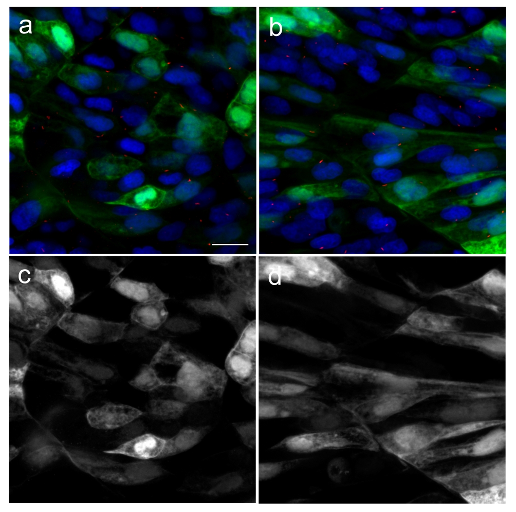
Figure Caption
Fig. S1 Green fluorescent protein (GFP) does not localize to primary cilia. In order to confirm that GFP alone does not localize to the primary cilia, we cloned GFP into pCS2 vector and injected into newly fertilised zebrafish eggs. Injected embryos were fixed at 18 hpf. Primary cilia and nuclei were visualised with acetylated tubulin (red) and DAPI (blue) respectively. GFP can be seen to localize to the cytoplasm and nuclei in both slow (b) and fast muscle (a) cells, but was not detected in the primary cilia. Green channels from (a) and (b) are shown in (c) and (d), respectively. Scale bar:10 μm
Acknowledgments
This image is the copyrighted work of the attributed author or publisher, and
ZFIN has permission only to display this image to its users.
Additional permissions should be obtained from the applicable author or publisher of the image.
Open Access.
Full text @ BMC Biol.