
|
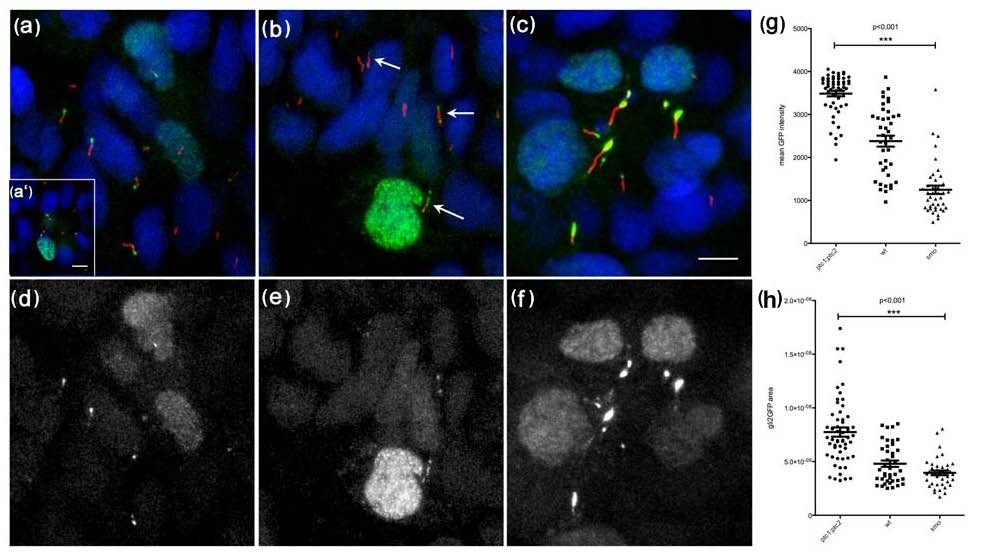
Fig. 2 Gli2a-GFP localizes to the distal tips of primary cilia in paraxial mesodermal cells (18 somite stage) and its localization is modulated by the activity of Hedgehog signalling. Gli2a-GFP injected embryos were labelled with mAb acetylated tubulin (red in a, b and c) and gamma tubulin (red in a′). This revealed that Gli2a-GFP was localized at the distal tip of primary cilia (a) and that its expression was excluded from the basal bodies (a′). In smoothened (smo) mutant embryos, levels of Gli2a-GFP were diminished (b) and the signal was dispersed along the cilia or localized both at the distal tip and at the basal bodies (arrows). In ptc1:ptc2 double mutant embryos, by contrast, high levels of Gli2a-GFP accumulated at the distal tip of the cilia (c). Panels (d-f) show the green channel images of (a-c), respectively. The intensity and the area of Gli2a-GFP at the tip of the cilia were measured in wild type and mutant embryos and the difference between the three groups was analysed by one way ANOVA test. This revealed a significant difference in protein levels between wild type, smo and ptc1;ptc2 mutant groups with P < 0.001 (g and h). Scale bars: 5 μm