
|
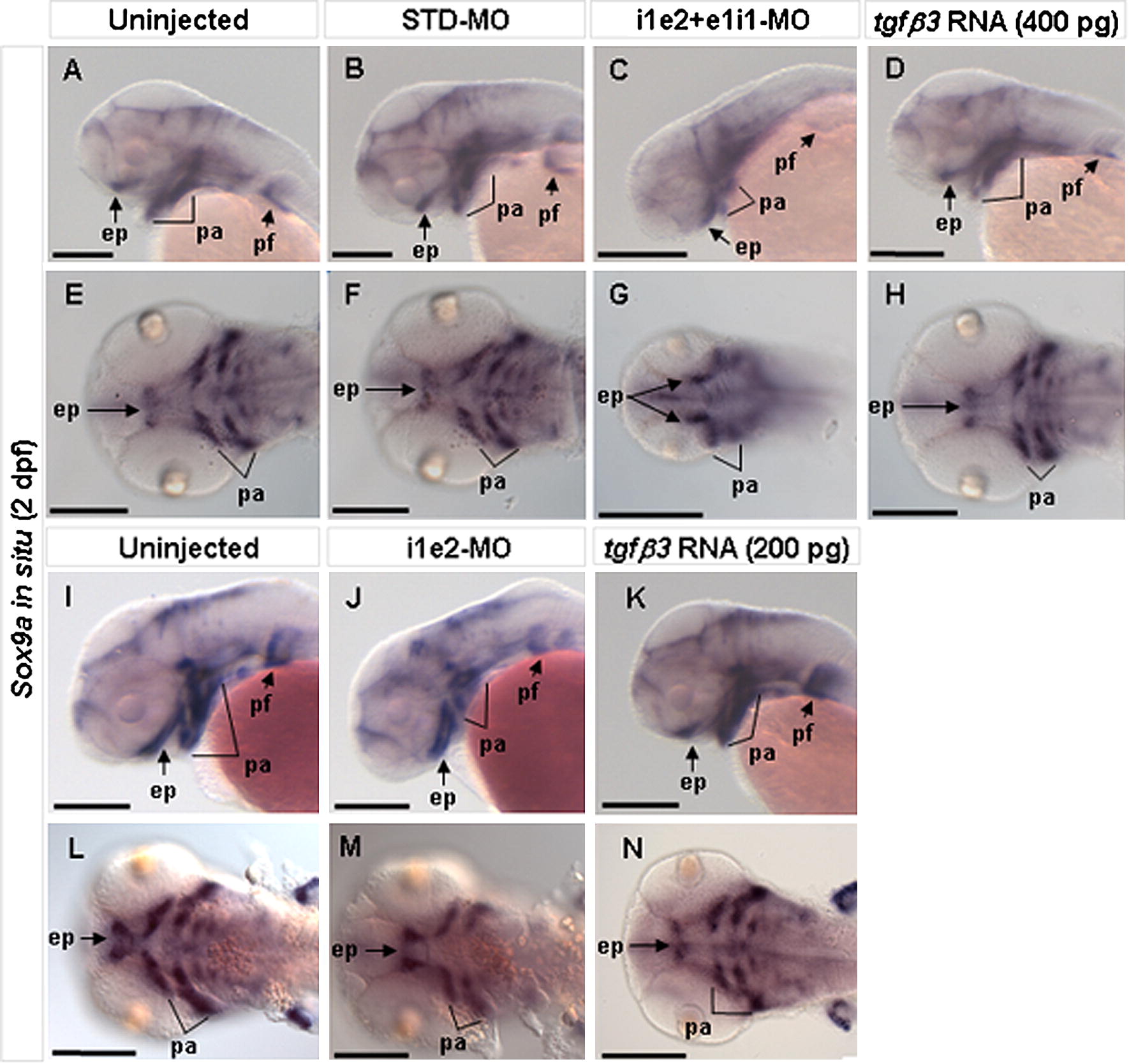
Fig. 3 Effect of tgfβ3 knockdown and over-expression on craniofacial chondrogenesis in 2 dpf embryos (A–N). Whole-mount lateral (A–D, I–K), and flat-mount ventral (E–H, L–N) views are shown. Expression of the cartilage differentiation marker sox9a appeared similar in uninjected and STD-MO injected embryos (A, B, E, F, I, L). In the i1e2 + e1i1 double morphant, however, there is markedly reduced sox9a expression in the developing pharyngeal arches and absent expression in the medial ethmoid plate (C, G). Sox9a expression is also mildly reduced in tgfβ3 over-expressing embryos (400 pg mRNA), with absent expression in the medial ethmoid plate (D, H). In the i1e2 single morphant, there is still a reduced sox9a expression in the developing pharyngeal arches and absent expression in the medial ethmoid plate (J, M) compared to the uninjected control embryos (I, L). Similarly, sox9a expression is also weakly reduced in 200 pg tgfβ3 mRNA injected embryos (K, N). ep, ethmoid plate; pa, pharyngeal arches; pf, pectoral fin. Black scale bars, 200 μm.
Reprinted from Mechanisms of Development, 128(7-8), Cheah, F.S., Winkler, C., Jabs, E.W., and Chong, S.S., tgfbeta3 Regulation of Chondrogenesis and Osteogenesis in Zebrafish is Mediated Through Formation and Survival of a Subpopulation of the Cranial Neural Crest, 329-344, Copyright (2010) with permission from Elsevier. Full text @ Mech. Dev.