
|
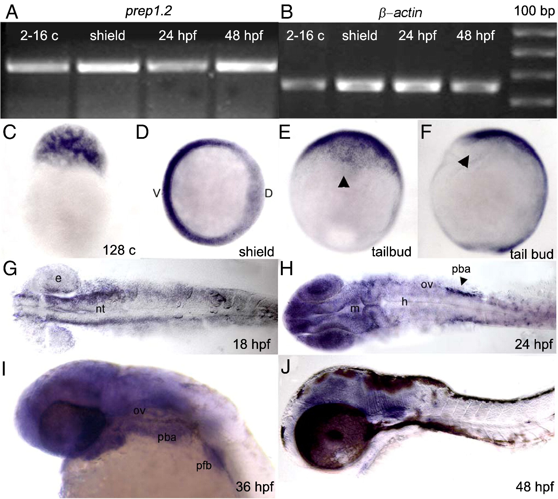
Fig. 1 Expression of prep1.2 during development. A, B: RT-PCR analysis showing amplification of prep1.2 from morula to 48 hpf stages. C–J: Whole-mount in situ hybridization experiments with a prep1.2 antisense probe showing that prep1.2 is weakly and ubiquitously expressed from 128 cells to tailbud stage (panels from C to F). G: during somitogenesis prep1.2 becomes more concentrated in cells localized on each side of the neural tube. H: At 24 hpf prep1.2 transcripts remain weakly ubiquitous with stronger expression in the CNS (forebrain and midbrain), eyes and in cell clusters localized on each side of the NS, posterior to the otic vesicle. I: at 36 hpf prep1.2 is expressed in the head and in the pharyngeal and pectoral fin bud mesenchyme. J: From 48 hpf onwards prep1.2 expression decreases and is restricted to the head region. e: eye; h: hindbrain; m: midbrain; nt: neural tube; ov: otic vesicle; pba: prospective branchial arches; pfb: pectoral fin bud. D: Top view with ventral side to the left. E: Frontal view. F: lateral view. G and H: Embryos are in dorsal view. I and J: Embryos are in lateral view.
Reprinted from Developmental Biology, 343(1-2), Vaccari, E., Deflorian, G., Bernardi, E., Pauls, S., Tiso, N., Bortolussi, M., and Argenton, F., prep1.2 and aldh1a2 participate to a positive loop required for branchial arches development in zebrafish, 94-103, Copyright (2010) with permission from Elsevier. Full text @ Dev. Biol.