
Image
|
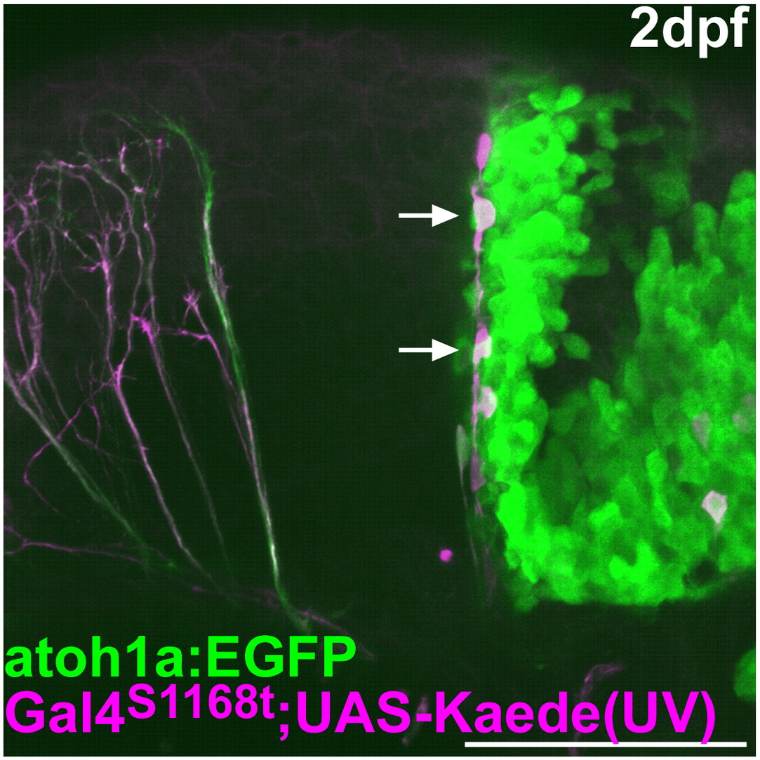
Figure Caption
Fig. S3 Lineage tracing of the rostro-medial atoh1a+ cells. As described in Fig. 2, the green Kaede in the rostral cerebellum was converted to a red fluorescent protein (magenta) by UV irradiation at 24 hpf in Et(fos:Gal4-VP16)s1168t; Tg(UAS-E1b: Kaede)s1999t; Tg(atoh1a:EGFP). The atoh1a:EGFP and the red Kaede were detected at 2 dpf. Lateral view with rostral to the left. The cells migrating ventro-laterally from the rostro-medial cerebellum are marked by arrows. Scale bar: 100 μm.
Acknowledgments
This image is the copyrighted work of the attributed author or publisher, and
ZFIN has permission only to display this image to its users.
Additional permissions should be obtained from the applicable author or publisher of the image.
Reprinted from Developmental Biology, 343(1-2), Kani, S., Bae, Y.K., Shimizu, T., Tanabe, K., Satou, C., Parsons, M.J., Scott, E., Higashijima, S.I., and Hibi, M., Proneural gene-linked neurogenesis in zebrafish cerebellum, 1-17, Copyright (2010) with permission from Elsevier. Full text @ Dev. Biol.