
|
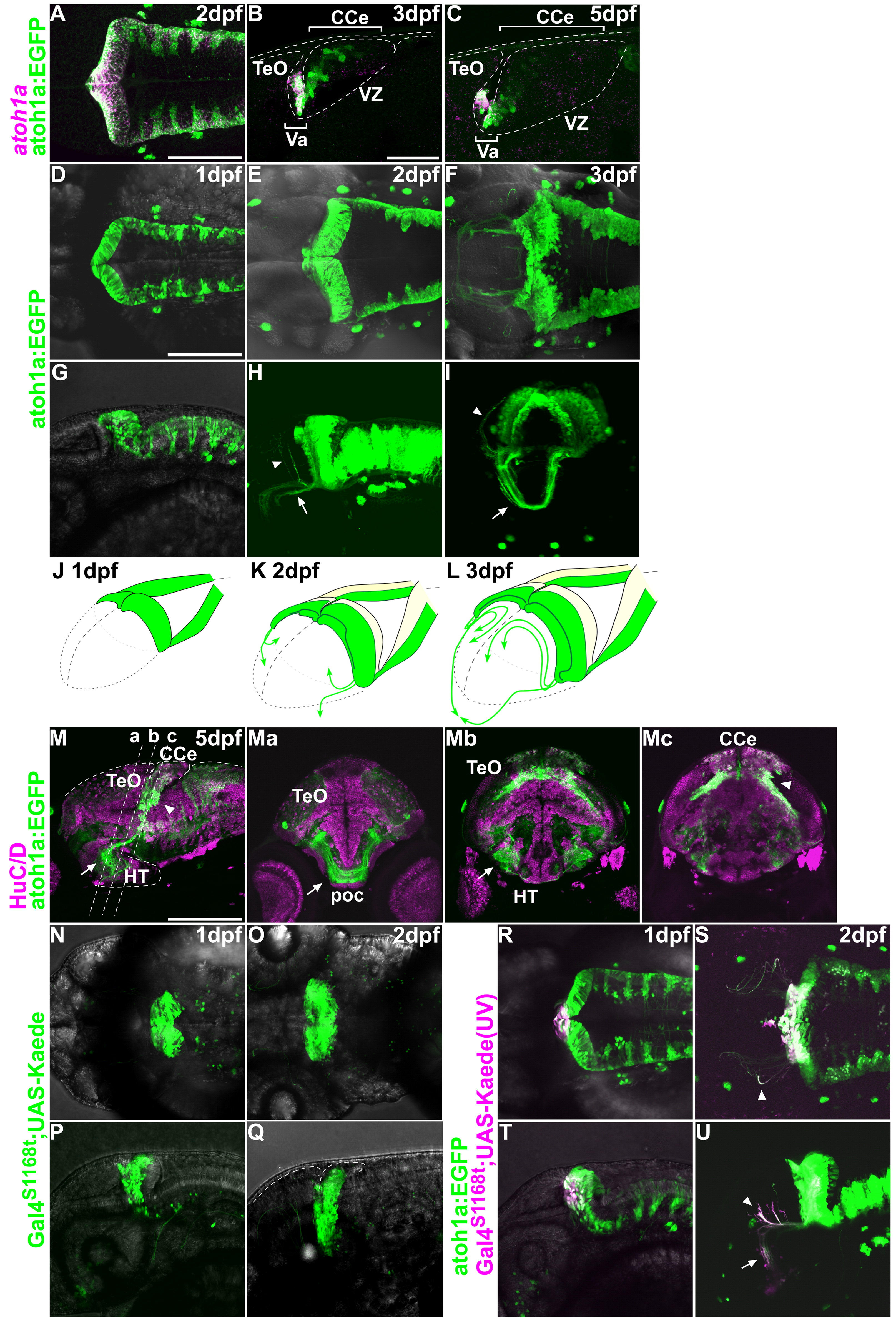
Fig. 2 Behaviors of neurons derived from atoh1a+ neuronal progenitors. (A–C) Expression of atoh1a mRNA (magenta) and EGFP (atoh1a:EGFP, green) in the cerebellum of Tg(atoh1a:EGFP) fish at 2, 3, and 5 dpf. Dorsal view (A) and parasagittal sections (B, C), with rostral to the left. Note that the atoh1a:EGFP signals overlap those of the atoh1a transcripts at 2 dpf in the valvula cerebelli (Va) but not the corpus cerebelli (CCe). The atoh1a-, atoh1a:EGFP+ cells are descendants of atoh1a+ progenitors. (D–J) Expression of atoh1a:EGFP at 1 (D, G), 2 (E, H), and 3 dpf (F, I). Dorsal (D–F), lateral (G, H), antero-lateral (I) views. Arrows and arrowheads indicate axonal projections to the optic tectum and hypothalamus, respectively (H, I). (J-L) Schematic representation of atoh1:EGFP+ cells and their projections at 1 (J), 2 (K), and 3 dpf (L). (M) Expression of atoh1a:EGFP (green) and HuC/D (magenta) at 5 dpf. Sagittal section (M). (Ma, b, c) Cross sections at the levels indicated in (M). Arrows indicate tegmental nuclei derived from the URL, which include secondary gustatory nucleus (SGN). Projections from the atoh1a:EGFP+ SGN are indicated by arrowheads. (N–U) Lineage tracing of the rostro-medial atoh1a+ cells using the photoconvertible protein Kaede. Green (non-photoconverted) Kaede was detected in the rostral cerebellum in Et(fos:Gal4-VP16)s1168t; Tg(UAS-E1b: Kaede)s1999t at 1 (N, P) and 2 dpf (O, Q). The tectum and cerebellum are indicated by dotted lines (Q). The green fluorescent Kaede in the rostro-medial cerebellum was converted to a red fluorescent protein (magenta) by UV irradiation at 24 hpf in Et(fos:Gal4-VP16)s1168t; Tg(UAS-E1b: Kaede)s1999t; Tg(atoh1a:EGFP) (R-U). The Kaede expression overlapped with that of atoh1a:EGFP in the rostro-medial cerebellum (R, T, and Suppl. Fig. 2). These rostro-medial cells migrated ventro-laterally (S, U, and Suppl. Fig. 3) were located at the bottom of the cerebellum, and projected to the optic tectum (arrowheads) and hypothalamus (arrow). These cells were located in the tegmentum at 5 dpf (M). HT, hypothalamus; poc, postoptic commissure. Other abbreviations are the same as in Fig. 1. Scale bars: 100 μm (B, D), 200 μm (M).
Reprinted from Developmental Biology, 343(1-2), Kani, S., Bae, Y.K., Shimizu, T., Tanabe, K., Satou, C., Parsons, M.J., Scott, E., Higashijima, S.I., and Hibi, M., Proneural gene-linked neurogenesis in zebrafish cerebellum, 1-17, Copyright (2010) with permission from Elsevier. Full text @ Dev. Biol.