
|
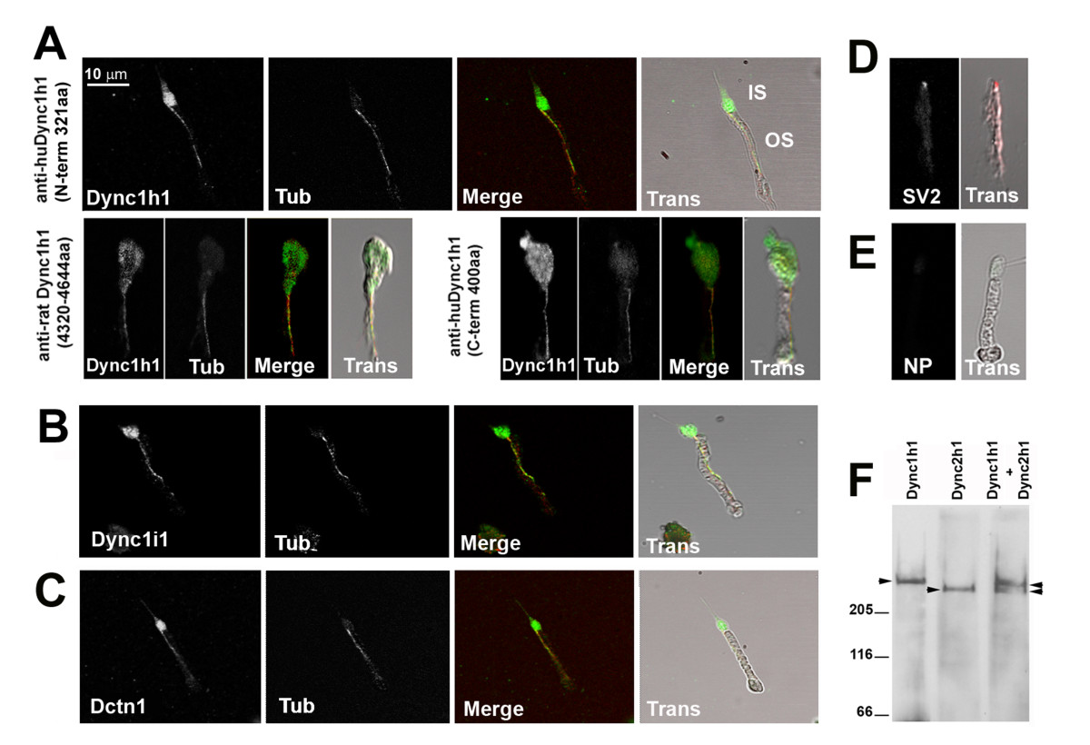
Fig. 7 Dync1h1, Dync1i1, and Dnct1 immunoreactivity is associated with photoreceptor axonemes. Immunocytochemical localization of proteins in isolated adult zebrafish photoreceptor cells. (A) Immunocytochemical localization of Dynein1 heavy chain (Dync1h1) using antisera directed against three different epitopes. (B) Immunocytochemical localization of Dynein1 intermediate chain (Dync1i1), (C) Dynactin1/p150 (Dctn1), and (D) Synaptic vesicle protein 2 (SV2). (E) Immunostaining in which the primary antibody was omitted. In (A-C), cells were co-stained with acetylated α-tubulin (Tub) to highlight the axoneme. Color merged (Merge) and transmitted light images overlaid with Merge images (Trans) are shown for each panel. IS, inner segment; OS, outer segment. NP, no primary antibody. (F) Western blot analysis on detergent-extracted photoreceptor cytoskeleton (DEPC) fraction shows the specificity of the Dync1h1 antibody, as the Dync2 h2 antibody recognizes a discrete and faster migrating band.