
|
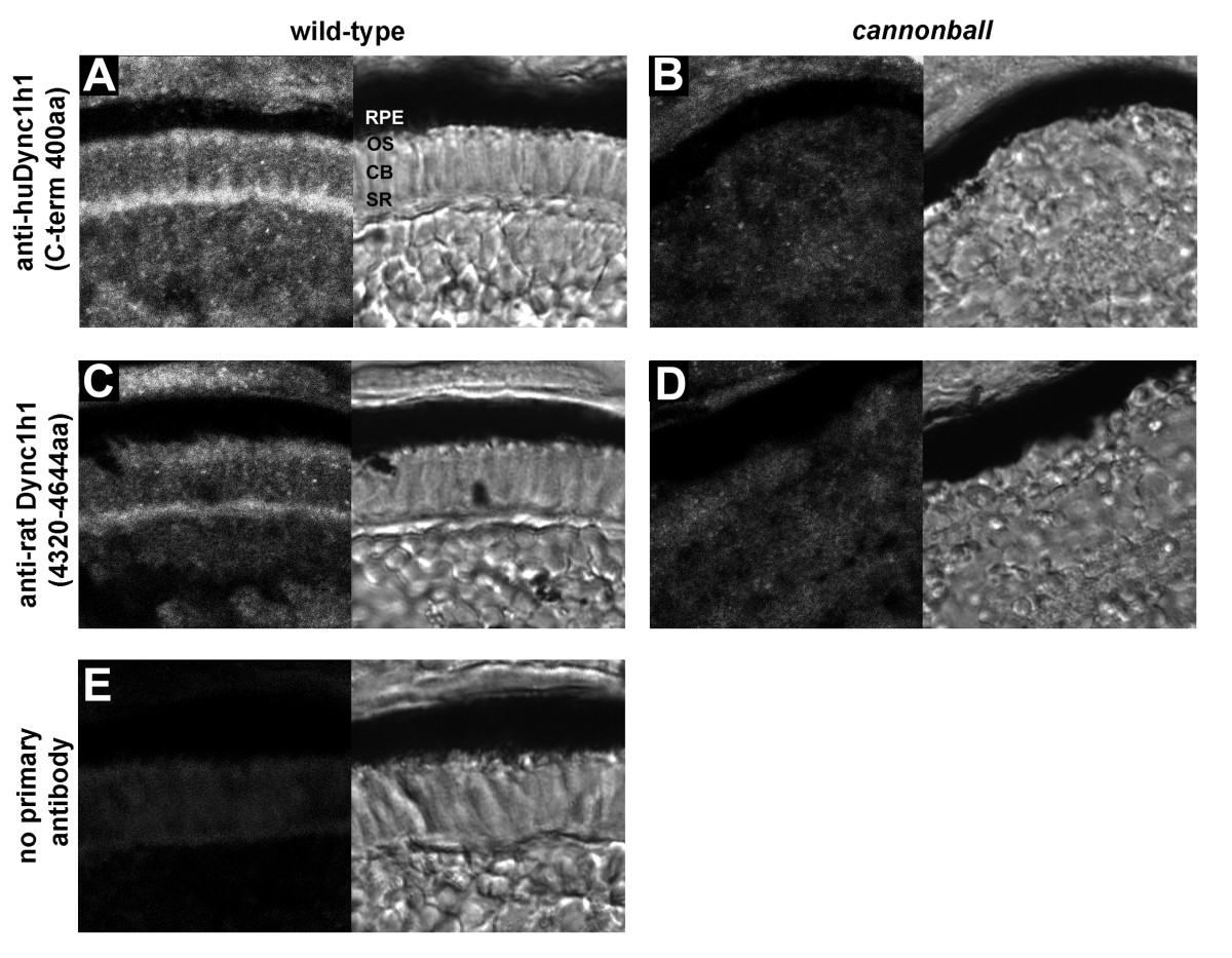
Fig. 6 Immunolocalization of Dync1h1 in developing photoreceptors. (A-E) Cryosections of the photoreceptor layer from wild-type retina (A, C, E) or cnb mutant retina (B, D) each at 4 dpf. Antisera directed against two different regions of Dync1h1 was used: (A, B) anti-human DYNC1H1 (huDYNC1H1; carboxy-terminal 400 amino acids) and (C, D) anti-rat Dync1h1 (amino acids 4,320 to 4,644). Primary antibodies were omitted in (E). For each panel, the fluorescent image is shown on the left and the transmitted light image on the right. CB, cell body; OS, outer segment; RPE, retina pigment epithelium; SR, synaptic region.