
|
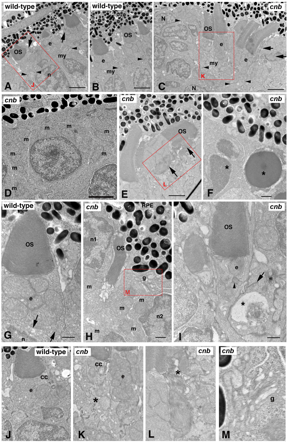
Fig. 5 The cnb mutation disrupts photoreceptor polarity and outer segment formation. (A, B) TEM of a wild-type central retina at 3 dpf showing normal structure of outer segments (OS) with aligned discs adjacent to the ellipsoid (e) region of the inner segments with bundles of tightly compacted mitochondria. The myoid (my) of the inner segment is the region between the mitochondria-rich ellipsoid and the elongated nuclei (n). Dense adherens junctions (arrowheads) can be seen at the external limiting membrane. Arrows in (A) point to connecting cilia. See also (J) for high magnification. (C-F, H, I) TEM of cnb mutant retina. (C) TEM of a cnb central retina at 3 dpf with both normal photoreceptor outer segments (left) and cells with disorganized outer segments (right, arrows). Adherens junctions are indicated by arrowheads. Overall, photoreceptors in the peripheral retina showed more severe defects, including cells that failed to form a polarized inner segment and cells with pyknotic nuclei. (D) Higher magnification of peripheral cnb photoreceptors that lack polarity and display dispersed mitochondria (m). (E) Normal cnb photoreceptors (left) next to an abnormal cell with a disrupted outer segment (arrows). See (L) for higher magnification. (F) Photoreceptors at the periphery with pyknotic nuclei (asterisks) and dense granular cytoplasm. (G) High magnification image of polarized wild-type cone photoreceptor at 3 dpf showing normal outer segment, typical ellipsoid with clustered mitochondria, and a narrow myoid with membrane vesicles and cisternae (arrows). (H, I) Central cone photoreceptors of cnb mutant eyes showing abnormal polarity. In (H), the cell on the left labeled n1 has its ellipsoidal mitochondria (m) on the opposite side of the nucleus from the retinal pigment epithelium (RPE) with an adjacent miss-aligned outer segment. The cell on the right (n2) has its Golgi complex (g) adjacent to RPE and the mitochondria (m) is scattered throughout the cytoplasm. A higher magnification of the Golgi is shown in (L). In (I), a cnb cone photoreceptor with a normal OS has a single mitochondrion at the position of the ellipsoid (e) and a displaced centriole (arrowhead). The adjacent cell on the right displays an abnormally positioned synaptic region with a large vacuole (asterisk) and a synaptic ribbon (arrow) adjacent to the inner segment of the cone cell in the middle. (J-M) Higher magnification insets of the red boxed regions highlighting the connecting cilium (cc) and ellipsoid (e) regions. Asterisk in (K) indicates a cell with a disorganized inner segment adjacent to a cell with a more normal ellipsoid. Asterisk in (L) indicates disorganized outer segment. Scale bars: 2 μm in (A-C); 0.5 μm in (F-I).