
|
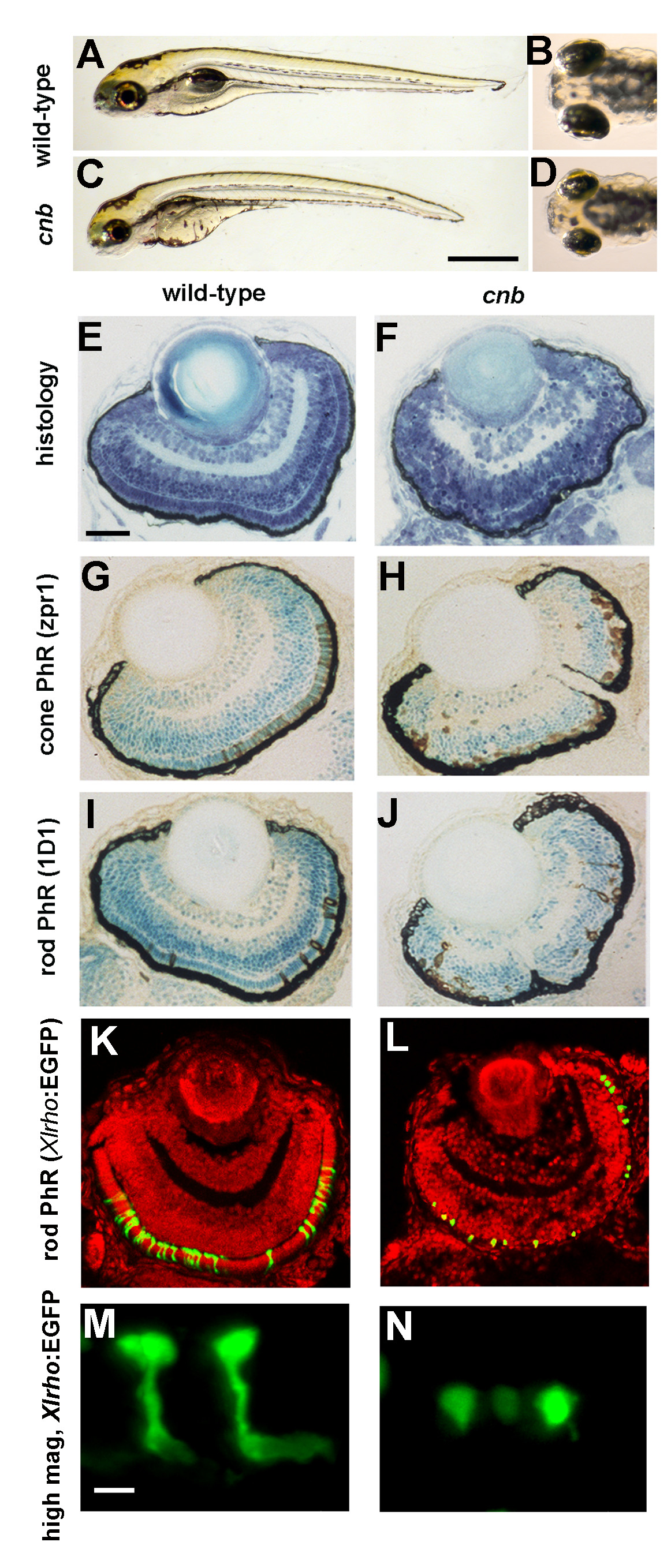
Fig. 1 The cnb mutation disrupts photoreceptor development. (A-D) Side (A, C) and dorsal (B, D) views of 5-dpf wild-type (A, B) and cnb mutant (C, D) embryos. (E-F) Retinal histology of wild-type (E) and cnb (F) retina at 4 dpf. (G, H) Zpr-1 immunoreactivity (brown) to label double cone photoreceptors (PhR) in 4-dpf wild-type (G) and cnb embryos (H). (I, J) ID1 immunoreactivity (brown) to label rod photoreceptors in 4-dpf wild-type (G) and cnb embryos (H). Nuclei in (G, H) are counterstained with methylene blue. (K-N) Confocal microscopy of wild-type (K, M) and cnb (L, N) whole retina (K, L) and individual rod photoreceptor cells (M, N) from Tg(Xlrho:EGFP)fl1 fish. Green, Xlrho:EGFP-positive cells; red, propidium iodide positive nuclei. Scale bars: 1 mm in (A, C); 40 μm in (E-L); 5 μm in (M, N). EGFP, enhanced green fluorescent protein.