
|
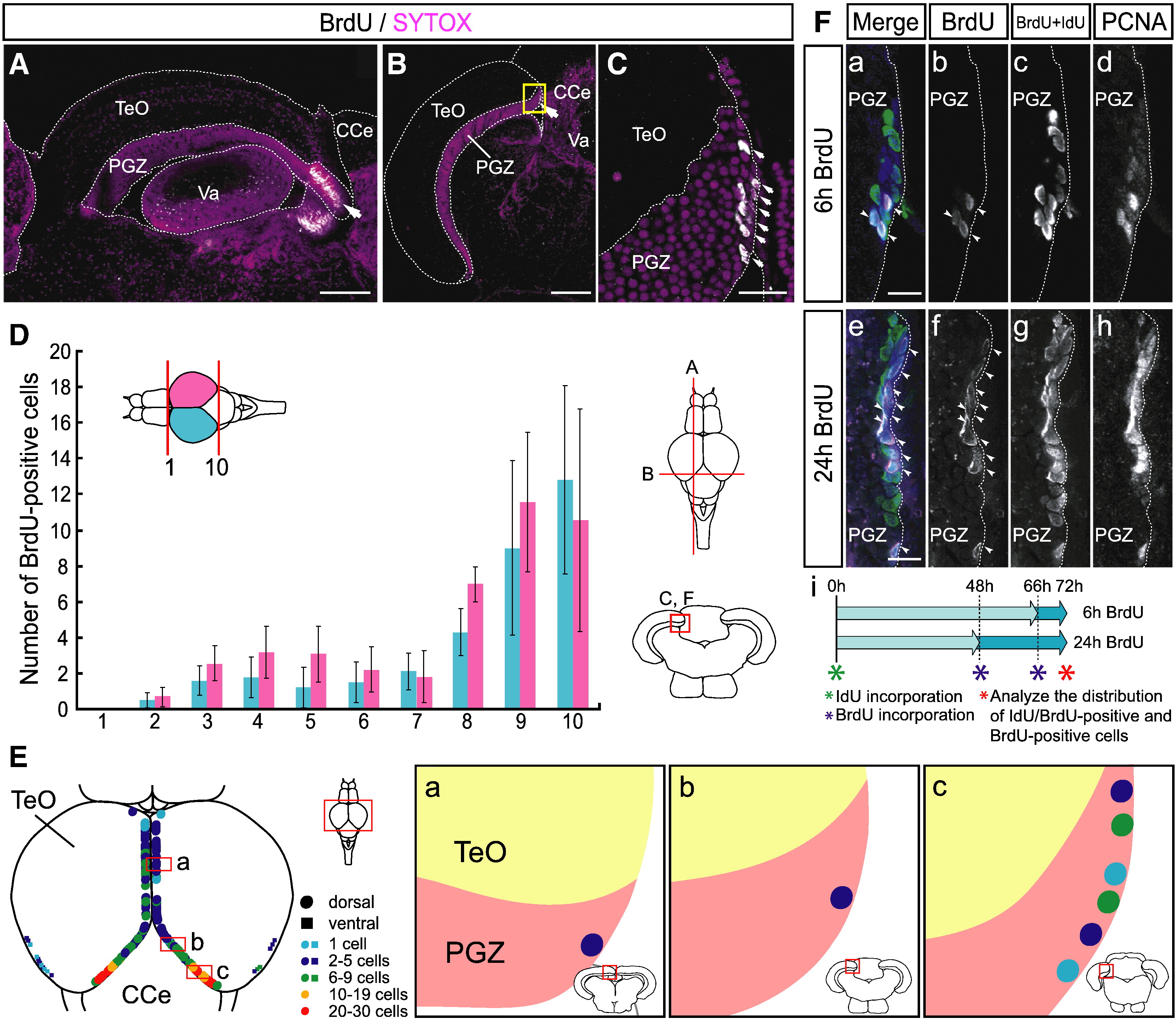
Fig. 1 Proliferating cells are distributed through the dorsomedial area of the caudal region of the PGZ in the adult zebrafish optic tectum. (A–C) Proliferating cells in the adult zebrafish optic tectum. Proliferating cells are labeled by 72-hour BrdU administration (white). Cell Nuclei are stained by SYTOX orange (red). (A) Sagittal section of adult zebrafish optic tectum (single plane, anterior left). A large cluster of BrdU-positive proliferating cells are distributed in the caudal region of the PGZ (arrow). (B) Transverse section of the caudal region of the adult zebrafish optic tectum (single plane, dorsal top). BrdU-positive proliferating cells are distributed through the dorsomedial area of the PGZ (arrow). (C) A high magnification view of the dorsomedial area of the PGZ, indicated by the yellow box in B (single plane, dorsal top). BrdU-positive proliferating cells are located on the dorsomedial margin (arrows). (D) Quantitative data of the distribution of BrdU-positive proliferating cells in the PGZ of the adult zebrafish optic tectum. The whole tectal region is divided into 10 parts along the rostrocaudal axis. Large numbers of proliferating cells are distributed through the caudal region of the optic tectum PGZ. Data are expressed as means ± SEM; n = 3. (E) Schematic drawing of the distribution of proliferating cells in the adult zebrafish optic tectum (dorsal view, anterior top, (a–c) transverse view, dorsal top). Proliferating cells are located along the dorsomedial and ventrolateral margins of the PGZ. The majority of BrdU-positive proliferating cells reside in the dorsomedial area of the PGZ caudal region (c). (F) Dorsomedial distribution of proliferating cells after 6 h (a–d) and 24 h of (e–h) BrdU labeling. One-third and one-half of the proliferating cells are labeled by 6 h and 24 h of BrdU administration, respectively (a, b, e, f, arrowheads). Most of the PCNA-positive cells are labeled by 72 h of continuous administration of IdU or BrdU, and visualized by BrdU antibody which detects both BrdU and IdU (c, g). Whole population of proliferating cells as visualized by immunohistochemistry with anti-PCNA antibody (d, h). IdU and BrdU double-labeling scheme is illustrated in panel i. CCe, corpus cerebelli; PGZ, periventricular gray zone; Tel, telencephalon; TeO, tectum opticum; Va, valvula cerebelli. Scale bars: 200 μm in A, B; 20 μm in C; 10 μm in F.
Reprinted from Developmental Biology, 342(1), Ito, Y., Tanaka, H., Okamoto, H., and Ohshima, T., Characterization of neural stem cells and their progeny in the adult zebrafish optic tectum, 26-38, Copyright (2010) with permission from Elsevier. Full text @ Dev. Biol.