
Image
|
Figure Caption
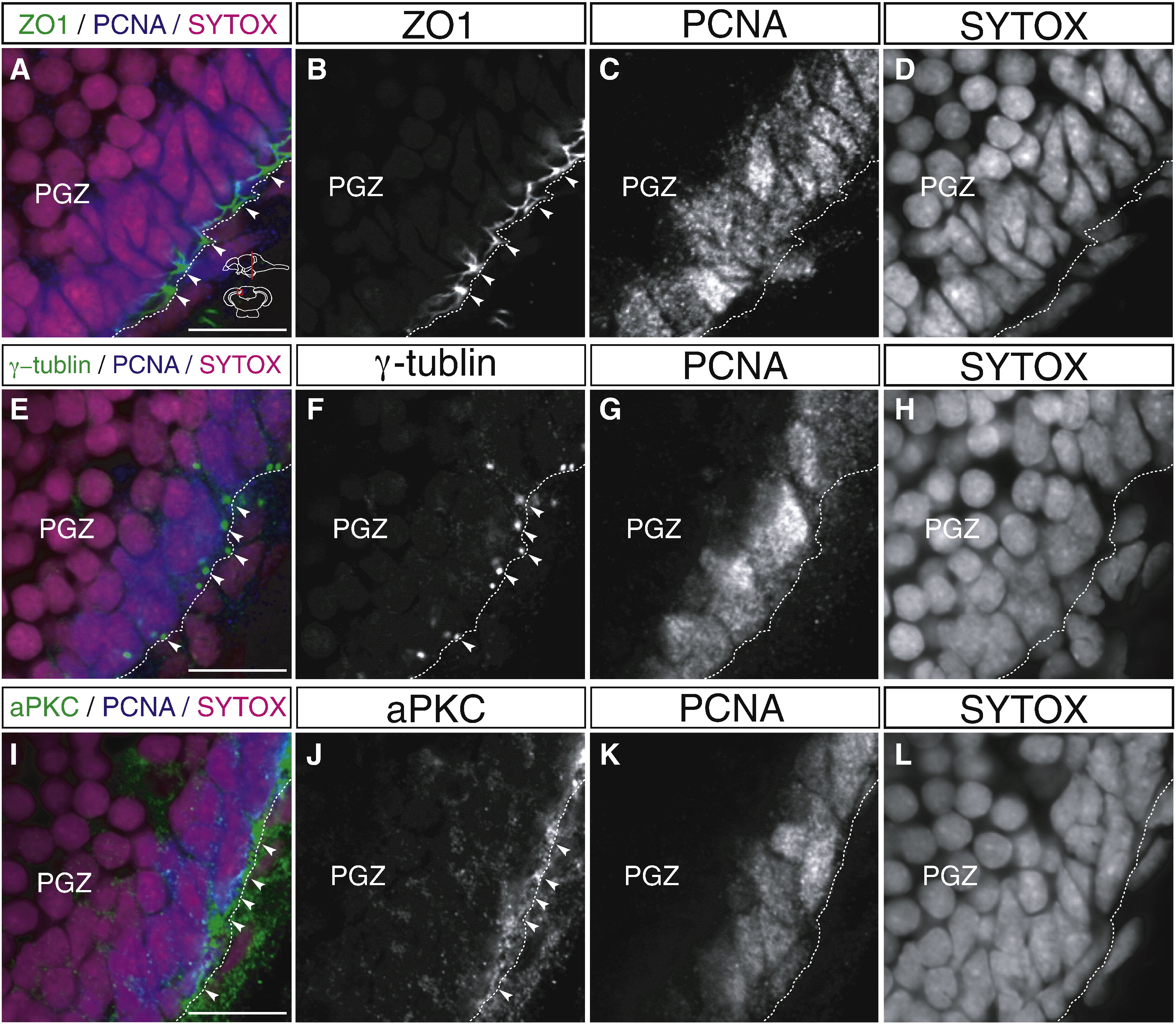
Fig. 5 Proliferating cells which face the ventricle maintain apical–basal polarity. (A–L) Localization of apical markers, ZO1 (A–D), γ-tubulin (E–H), and aPKC (I–L) in the proliferating cells of the PGZ of the adult zebrafish optic tectum (60 μm transverse sections, single planes, dorsal top). The proliferating cells are visualized by immunohistochemistry with anti-PCNA antibody. The proliferating cells which localize near the ventricle show highly polarized expression of apical markers such as ZO1 (A, B, arrowheads), γ-tubulin (E, F, arrowheads), and aPKC (I, J, arrowheads). Scale bars: 10 μm.
Acknowledgments
This image is the copyrighted work of the attributed author or publisher, and
ZFIN has permission only to display this image to its users.
Additional permissions should be obtained from the applicable author or publisher of the image.
Reprinted from Developmental Biology, 342(1), Ito, Y., Tanaka, H., Okamoto, H., and Ohshima, T., Characterization of neural stem cells and their progeny in the adult zebrafish optic tectum, 26-38, Copyright (2010) with permission from Elsevier. Full text @ Dev. Biol.