
|
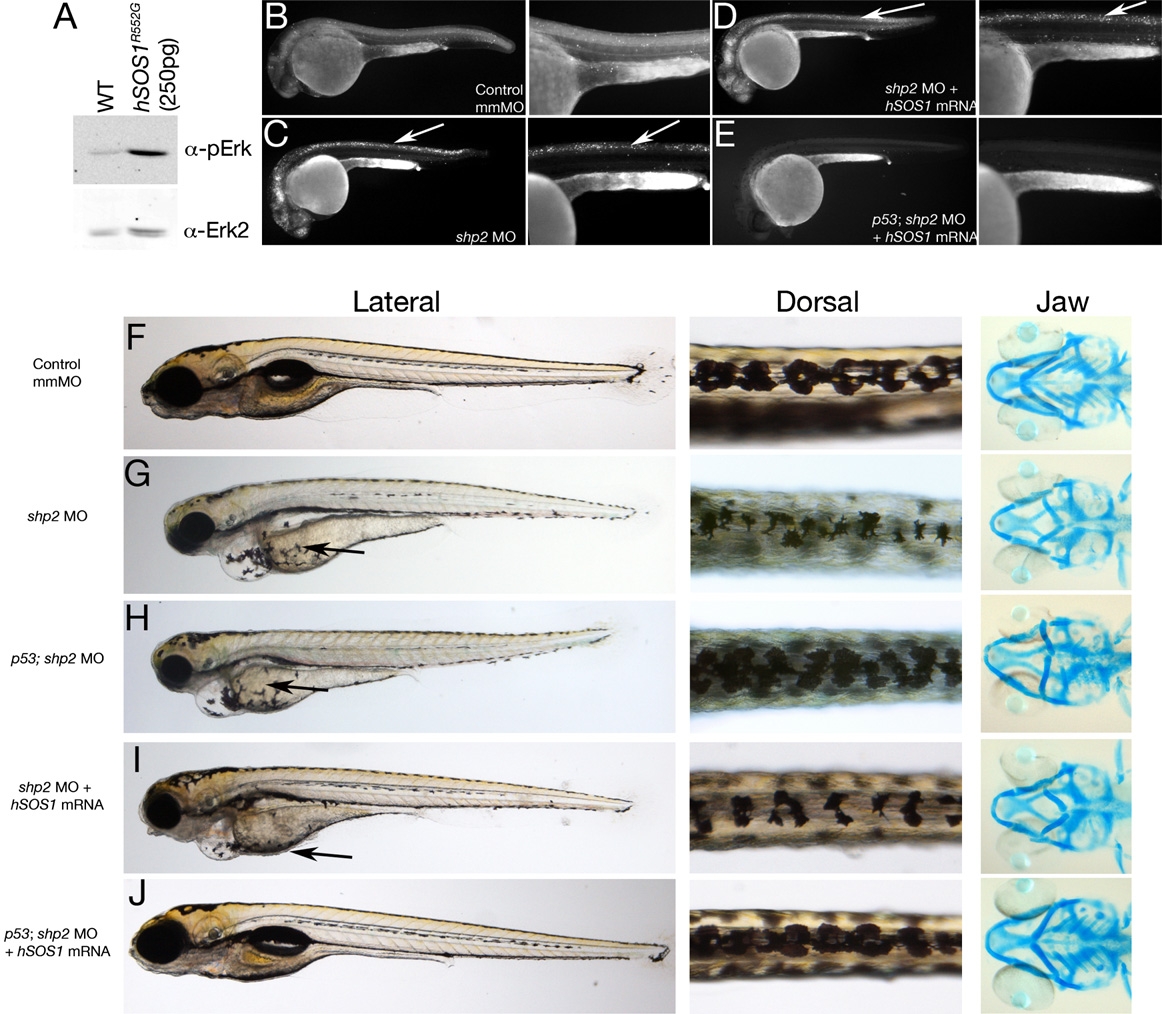
Fig. 5 Restoring Both Shp2-Dependent Pathways in shp2 MO Embryos Rescues Neural Crest Phenotypes
(A) Immunoblot showing effect of hSOS1R552G mRNA on Erk activation.
(B–E) Lateral views of Acridine orange labeling in 24 hpf embryos. Right panels in each pair are higher magnification views. Cell death caused by shp2 MO (arrows in [C]) is not rescued by coexpression with hSOS1R552G mRNA (arrows in [D]) in WT embryos, but is rescued in p53 mutant embryos (E).
(F–J) Lateral (left) and dorsal (middle) views of live 5 dpf embryos and ventral views (right) of 6 dpf embryos stained with Alcian blue. (G) Injection of shp2 MO causes severe jaw, heart, and pigmentation defects and delayed cell migration (arrow). (H) Injection of the shp2 MO into p53 mutant embryos partially rescues the jaw defects, but restores pigment cell numbers, although migration is still delayed (arrows in [G] and [H]). (I) Migration of pigment cells to the ventral stripe is rescued in shp2 MO embryos coinjected with hSOS1R552G mRNA (arrow), but pigment cell numbers are still reduced, particularly along the dorsal stripe (arrowheads), and embryos have smaller heads, heart edema, and severe craniofacial defects. (J) Coinjection of hSOS1R552G mRNA into p53 mutant embryos completely rescues neural crest phenotypes.
Reprinted from Developmental Cell, 18(5), Stewart, R.A., Sanda, T., Widlund, H.R., Zhu, S., Swanson, K.D., Hurley, A.D., Bentires-Alj, M., Fisher, D.E., Kontaridis, M.I., Look, A.T., and Neel, B.G., Phosphatase-Dependent and -Independent Functions of Shp2 in Neural Crest Cells Underlie LEOPARD Syndrome Pathogenesis, 750-762, Copyright (2010) with permission from Elsevier. Full text @ Dev. Cell